Journal List > Yonsei Med J > v.47(2) > 1029985

Song, Song, Choi, Kim, and Rhee: Plasma Level of IL-6 and Its Relationship to Procoagulant and Fibrinolytic Markers in Acute Ischemic Stroke

Abstract

Procoagulant or impaired fibrinolytic states as well as inflammatory reactions mediated by cytokines are likely involved in the pathogenesis of acute ischemic stroke. We examined the potential relationship between interleukin 6 (IL-6) and hemostatic markers. The procoagulant and fibrinolytic states were assessed in 46 patients with acute stroke by measuring plasma levels of plasminogen activator inhibitor-1 (PAI-1), thrombin-antithrombin complex (TAT), and plasminogen-antiplasmin complex (PAP). Circulating IL-6 levels were measured using ELISA (Quantikine, R&D systems, MN, USA). Circulating IL-6 (mean, 26.5 pg/mL) and PAI-1 (mean, 19.9 ng/mL) levels were higher in patients with acute stroke than in healthy subjects (mean, 3.0 pg/mL, 10.4 ng/mL, respectively). TAT levels were statistically different according to the etiologic subtypes of stroke (atherogenic, 2.5 ng/mL; lacunar 3.2 ng/mL; cardiogenic 9.9 ng/mL, p = 0.021). Neither procoagulant levels nor fibrinolytic markers significantly correlated with circulating IL-6 levels. Our findings suggest that elevated proinflammatory cytokines during the initial hours of ischemic stroke may be an independent pathogenic factor or a consequence of the thrombotic event with no relationship to the procoagulant or fibrinolytic states.

INTRODUCTION

Atherosclerosis is the main cause of cerebral infarction.1 The revised response to injury hypothesis highlights the importance of inflammatory reactions involved in the pathogenesis of atherosclerosis.2 Injury of endothelial cells may alter the balance of procoagulant and antithrombotic factors on the surface of endothelium to favor thrombosis.3 Consonant with the role of monocytes/macrophages, vascular smooth muscle cells and platelets, there are several important interactions between the blood components of both the coagulation and fibrinolytic systems.4 Thus, a growing body of evidence supports a relationship between the inflammatory response, thrombosis, and the development of atherosclerotic lesions.5 In addition, monocyte-derived cytokines, such as IL-6, are in part responsible for the elevations of plasma fibrinogen that accompany inflammation. To better understand the relationship between the inflammatory response and thrombosis in ischemic stroke, we assessed procoagulant and fibrinolytic states in patients with acute ischemic stroke by measuring plasma levels of plasminogen activator inhibitor-1 (PAI-1), thrombin-antithrombin complex (TAT) and plasminogen-antiplasmin complex (PAP), and examined the potential relationship between these hemostatic markers and interleukin 6 (IL-6).

MATERIALS AND METHODS

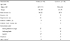
Forty-six patients (34 men and 12 women, mean age, 64 yrs) presenting with acute ischemic stroke were studied according to a standard protocol entailing clinical and imaging studies (CT, MRI and/or angiography) of the brain. Patients were admitted with atherosclerotic (n = 28), lacunar (n = 14), and cardioembolic (n = 4) infarcts within the first 24 hours, and 28 age- and sex- matched healthy subjects were included for comparison (Table 1). Citrated blood was drawn early in the morning on admission to determine plasma levels of TAT (Dade-Behring, Germany), PAP (Dade-Behring, Germany), PAI-1 (Diagnostica Stago, France), IL-6 (Quantikine, R&D systems, MN, USA), and anti-phospholipid antibody (IgG/IgM Diagnostica Stago, France) by ELISA methods.

RESULTS

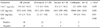
Circulating IL-6 levels (mean, 26.5 pg/mL, range, 6.4-161.3 pg/mL) were significantly higher in all patients with acute stroke than in healthy subjects (median, 3.0 pg/mL, range, 2.3-5.9 pg/mL) (p < 0.001, Table 2).
PAI-1 and PAP levels were also significantly different between patients with acute stroke (mean, 19.9 ng/mL, 522.9 ng/mL, respectively) and healthy subjects (mean 10.4 ng/mL, 240.4 ng/mL, respectively) (p < 0.001, Table 2).
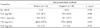
Mean levels of TAT (3.4 ng/mL) in the stroke patient group were not significantly increased compared with normal subjects (2.8 ng/mL) (p < 0.001, Table 2), however, TAT levels were statistically different according to the etiologic subtypes of stroke (atherogenic, 2.5 ng/mL; lacunar 3.2 ng/mL; cardiogenic, 9.9 mg/mL; p = 0.021).
PAI-1 and PAP levels were higher in the lacunar subtype and in the cardiogenic subtype, respectively, than in other subtypes, with weak statistical significance (p = 0.070 and p = 0.058, respectively). However, there were no significant differences in levels of IL-6 and other hemostatic markers among the different types of stroke (Table 3).
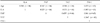
Levels of IL-6 and hemostatic parameters were not different between positive and negative groups for anti-phospholipid antibodies (Table 4).
There were significant correlations between TAT and PAP markers (r = 0.455, p = 0.0015), and TAT correlated weakly with PAI-1 (r=0.264, p = 0.0787). However, neither procoagulant nor fibrinolytic markers were significantly correlated with circulating IL-6 levels (Table 5).

DISCUSSION

Elevated plasma fibrinogen is a recognized risk factor for stroke6 and elevations in fibrinogen may in fact be a surrogate for elevated IL-6, a cytokine which plays a critical role in regulating fibrinogen production.7 IL-6 levels have also been reported to be elevated in stroke patients,8-10 and the increase of IL-6 significantly correlated with increased brain lesion volumes and was associated with poor functional and neurological outcome.9 In addition to elevated IL-6 levels in patients, gene expression of interleukin-1β (IL-1β) and TNF-α in rats are elevated after cerebral infarction.11 IL-6 also regulates the production of tumor necrosis factor (TNF-α),12 which in turn is capable of inducing tissue factor-like procoagulant activity on the surface of endothelial cells.13 In addition, these mediators activate the hepatic synthesis of acute-phase reactants such as C-reactive protein, serum amyloid A protein, fibrinogen, haptoglobin, ceruloplasmin, and C4b-binding protein (C4b-BP).11,14 C4b-BP possesses a binding site for protein S, a cofactor for protein C, and another natural anticoagulant which inactivates factors Va and VIIIa.15 In fact, during stroke, C4b-BP causes a decrease of free protein S,16 which may promote a procoagulant state. As a result, IL-6 may influence the disease course in the stroke by changing coagulation and fibrinolysis, in addition to causing hemorheologic changes by increasing fibrinogen levels and blood viscosity.17 Therefore, the aim of this study was to assess the relationship between circulating levels of IL-6 and hemostatic status in acute ischemic stroke.
In our study, IL-6 levels were significantly elevated early after stroke. This observation is consistent with previous studies8-10 which suggest that IL-6 may play an important role in the inflammatory changes that occur following brain ischemia and the subsequent tissue damage.
Despite a significant increase in IL-6 levels, circulating levels of TAT (a reflection of systemic procoagulant activity18) did not correlate with IL-6. This observation supports the hypothesis that IL-6 production is a localized inflammatory response to acute hypoxic-ischemic injury and is not related to enhanced thrombin activity or intravascular hypercoagulation status.
In this study we found that mean TAT levels shortly after stroke were mildly elevated (3.4 ng/mL), an observation similar to a previous report by Geiser et al. (2.9 ng/mL).19 These findings suggest that thrombin formation is only partially suppressed, despite adequate anticoagulation, in patients with cerebrovascular events with prosthetic heart valves. This is present in atherogenic or lacunar subtypes, suggesting increased procoagulant activity in patients with cardioembolism and different pathophysiology of each subtype. Even though there was no statistical difference in IL-6 levels among the three different subtypes, the mean value of IL-6 was highest in the cardiogenic type, an observation that needs to be studied further using a large number of patients of different subtypes.
Although the importance of the thrombotic process and fibrin metabolism in stroke has been recognized for years, the role of fibrinolytic abnormalities have not been clearly delineated, probably because of the heterogeneous nature of stroke syndrome and because of the variety of in vitro methods for assessing fibrinolysis.
The recent development of specific immunoassays including t-PA,20 PAI-1,21 and PAP22 allow more advanced assessment of the fibrinolytic system. In our study, IL-6 levels did not correlate with fibrinolytic markers, whereas there were significant correlations between procoagulant TAT and fibrinolytic markers (PAP and PAI-1). These findings reflect systemic derangements of both the clotting and fibrinolytic processes in stroke that do not involve IL-6.
The proinflammatory cytokines are produced not only by endothelial cells, monocytes, fibroblasts and T-lymphocytes,23,24 but also by microglia25 and astrocytes.26 Localized inflammatory response to acute brain lesion may have multiple local and systemic effects that modulate the disease course, including cytotoxicity,27 astrocyte proliferation,28 and leukocyte activation.29 Recently, Johansson et al.30 reported that neuroendocrine disturbances are often profound shortly after stroke, and cytokines including IL-6 seem to be important modulators of these disturbances. Vila et al.31 have also assessed the implication of IL-6 in early neurological worsening in ischemic stroke. More directly, IL-6 infusion has been shown to stimulate coagulation32 and to significantly correlate with coagulation markers.33 On the other hand, IL-6 levels during the acute phase should be interpreted with caution because the pattern of the related changes depends on the disease severity. Other contributing factors can also affect the coagulation system.34,35 Taken together, these results suggest that elevated proinflammatory cytokines during the initial hours of ischemic stroke may either be an independent pathogenetic factor or the consequence of the thrombotic event, with no relationship to the procoagulant or fibrinolytic states.
Further investigation into the role of IL-6 in stroke may provide a basis for new therapies of stoke prevention or treatment.

Figures and Tables

Table 1
Characteristics of Study Groups
ymj-47-201-i001

*p < 0.05 versus control group.

Other illness: chronic bronchitis, hyperthyroidism, multiple osteoarthritis, fatty liver, proteinuria, glaucoma, oral ulcer/gingivitis, degenerative spondylosis.

Table 2
Plasma Concentrations of IL-6 and PAI-1 in Patients with Acute Ischemic Stroke and Healthy Subjects
ymj-47-201-i002

Values are expressed as mean (range).

*Mann-Whitney test was used.

Data on PAI-1 were missing in one stroke patient.

Abbreviations: IL-6, interleukin-6; PAI, plasminogen activator inhibitor; TAT, thrombin-antithrombin; PAP, plasmin-antiplasmin.

Table 3
Circulating Levels of Hemostatic Parameters According to the Etiologic Subtypes of Acute Ischemic Stroke
ymj-47-201-i003

Values are given as number of subjects or mean (range).

*Chi-square test for categorical and ANOVA test for continuous variables were used.

Data on PAI-1 were missing in one cardiogenic subtypes.

Abbreviations: IL-6, interleukin-6; PAP, plasmin inhibitor complex; TAT, thrombin-antithrombin complex; PAI, plasminogen activator inhibitor; APA anti-phospholipid antibody (IgG of IgM).

Table 4
Circulating Levels of Cytokines and Hemostatic Parameters According to the Positivity (IgG or IgM) for Anti-phospholipid Antibody
ymj-47-201-i004

Values are given as number of subjects or mean values (SD).

*Chi-square test for categorical and Mann-Whitney test for continuous variables were used.

Data on PAI-1 were missing in 1 positive APA.

Abbreviations: see Table 2.

Table 5
Correlation Coefficients (r) Among Various Parameters in the Patients with Acute Ischemic Stroke
ymj-47-201-i005

*p = 0.0015, p = 0.0787 (Spearman correlation coefficients).

Abbreviations: see Table 2.

ACKNOWLEDGMENTS

The authors wish to thank Hae Sun Chun for her technical assistance in this study.

Notes

This study was supported by the Brain Korea 21 Project for Medical Science, Yonsei University, 2001.

References

1. Del Zoppo GJ, Zeumer H, Harker LA. Thrombolytic therapy in stroke : possibilities and hazards. Stroke. 1986. 17:595–607.
2. Tracy RP. Epidemiological evidence for inflammation in cardiovascular disease. Thromb Haemost. 1999. 82:826–831.
3. Scarpati EM, Sadler JE. Regulation of endothelial cell coagulant properties. Modulation of tissue factor, plasminogen activator inhibitors, and thrombomodulin by phorbol 12-myristate and tumor necrosis factor. J Biol Chem. 1989. 264:20705–20713.
4. Rabbani LE, Loscalzo J. Recent observations on the role of hemostatic determinants in the development of the atherothrombotic plaque. Atherosclerosis. 1994. 105:1–7.
5. Clark WM. Cytokines and reperfusion injury. Neurology. 1997. 49(5):Suppl 4. S10–S14.
6. Tsuda Y, Satoh K, Kitadai M, Takhashi T. Hemorheologic profiles of plasma fibrinogen and blood viscosity from silent to acute and chronic cerebral infarctions. J Neurol Sci. 1997. 147:49–54.
7. Castell JV, Andus T, Kunz D, Heinrich PC. Interleukin-6. The major regulator of acute phase protein synthesis in man and rat. Ann NY Acad Sci. 1989. 557:87–101.
8. Coull BM, Beamer NB, Clark WM. Elevated plasma interleukin-6 (IL-6) in acute stroke. Stroke. 1993. 24:183. (Abstract).
9. Fassbender K, Rossol S, Kammer T, Daffertshofer M, Wirth S, Dollman M, et al. Proinflammatory cytokines in serum of patients with acute cerebral ischemia: kinetics of secretion and relation to the extent of brain damage and outcome of disease. J Neurol Sci. 1994. 122:135–139.
10. Johansson A, Olsson T, Carlberg B, Karlsson K, Fagerlund M. Hypercorisolism after stroke-partly cytokine-mediated? J Neurol Sci. 1997. 147:43–47.
11. Vila N, Chamorro A. Cytokines and acute-phase response in acute stroke. Stroke. 1995. 26:1729–1731.
12. Remick DG, Kunkel RG, Larrick JW, Kunkel SL. Acute in vivo effects of human recombinant tumor necrosis factor. Lab Invest. 1987. 56:583–590.
13. Dobroski DR, Rabbani LE, Loscalzo J. Loscalzo J, Schfer AI, editors. The relationship between thrombosis and atherosclerosis. Thrombosis and Hemorrhage. 1998. Baltimore: Williams & Wilkins;837–861.
14. Marcovina SM, Zoppo A, Vigano-D'Angelo S, Di Cola G, D'Angelo A. Determination of serum levels of complement component C4b-binding protein: influence of age and inflammation. Int J Clin Lab Res. 1991. 21:171–175.
15. Esmon CT. The regulation of natural anticoagulant pathways. Science. 1987. 235:1348–1352.
16. Carr ME Jr, Zekert SL. Protein S and C4b-binding protein levels in patients with stroke: implications for protein S regulation. Haemostasis. 1993. 23:159–167.
17. Fisher M, Meiselman HJ. Hemorheological factors in cerebral ischemia. Stroke. 1991. 22:1164–1169.
18. Bick RL. Disseminated intravascular coagulation: pathophysiological mechanisms and manifestations. Semin Thromb Hemost. 1998. 24:3–18.
19. Geiser T, Sturzenegger M, Genewein U, Haeberli A, Beer JH. Mechanisms of cerebrovascular events as assessed by procoagulant activity, cerebral microemboli, and platelet microparticles in patients with prosthetic heart valves. Stroke. 1998. 29:1770–1777.
20. Feinberg WM, Bruck DC, Jeter MA, Corrigan JJ Jr. Fibrinolysis after acute ischemic stroke. Thromb Res. 1991. 64:117–127.
21. Takada A, Takada Y, Urano T, Sakakibara K, Shozuhara H, Toyama K. Changes in plasma levels of t-PA, PAI-1 and t-PA-PAI-1 complex in patients with cerebrovascular thrombosis infused with t-PA. Thromb Res. 1990. 59:375–381.
22. Bick RL, Murano G. Physiology of hemostasis. Clin Lab Med. 1994. 14:677–707.
23. Akira S, Hirano T, Taga T, Kishimoto T. Biology of multifunctional cytokines: IL 6 and related molecules (IL 1 and TNF). FASEB J. 1990. 4:2860–2867.
24. Libby P, Ordovas JM, Auger KR, Robbins AH, Birinyi LK, Dinarello CA. Endotoxin and tumor necrosis factor induce interleukin-1 gene expression in adult human vascular endothelial cells. Am J Pathol. 1986. 124:179–185.
25. Woodroofe MN, Sarna GS, Wadhwa M, Hayes GM, Loughlin AJ, Tinker A, et al. Detection of interleukin-1 and interleukin-6 in adult rat brain, following mechanical injury, by in vivo microdialysis: evidence of a role for microglia in cytokine production. J Neuroimmunol. 1991. 33:227–236.
26. Clark WM, Rinker LG, Lessov NS, Hazel K, Hill JK, Stenzel-Poore M, et al. Lack of interleukin-6 expression is not protective against focal central nervous system ischemia. Stroke. 2000. 31:1715–1720.
27. Araujo DM, Cotman CW. Transient neurotrophic effects of interleukins on hippocampal neurons in vitro (abstract). IIIrd IBRO World Congress of Neuroscience;243.
28. Giulian D, Lachman LB. Interleukin-1 stimulation of astroglial proliferation after brain injury. Science. 1985. 228:497–499.
29. Strieter RM, Kunkel SL, Showell HJ, Remick DG, Phan SH, Ward PA, et al. Endothelial cell gene expression of a neutrophil chemotactic factor by TNF-α, LPS, and IL-1β. Science. 1989. 243:1467–1469.
30. Johansson A, Ahren B, Nasman B, Carlstrom K, Olsson T. Cortisol axis abnormalities early after stroke--relationships to cytokines and leptin. J Intern Med. 2000. 247:179–187.
31. Vila N, Castillo J, Davalos A, Chamorro A. Proinflammatory cytokines and early neurological worsening in ischemic stroke. Stroke. 2000. 31:2325–2329.
32. Stouthard JM, Levi M, Hack CE, Veenhof CH, Romijin HA, Sauerwein HP, van der Poll T. Interleukin-6 stimulates coagulation, not fibrinolysis, in humans. Thromb Haemost. 1996. 76:738–742.
33. Aso Y, Okumura K, Yoshida N, Tayama K, Kinda T, Kobayashi I, et al. Plasma interleukin-6 is associated with coagulation in poorly controlled patients with type 2 diabetes. Diabet Med. 2003. 20:930–934.
34. Conrad MF, Stone DH, Albadawi H, Hua HT, Entabi F, Stoner MC, et al. Local inflammatory and thrombotic response differ in a murine model of partial and complete hindlimb ischemia/reperfusion. Surgery. 2005. 138:375–381.
35. Derhasching U, Bergmair D, Marsik C, Schlifke I, Wijdenes J, Jilma B. effect of interleukin-6 blockade on tissue fator-induced coagulation in human endotoxemia. Crit Care Med. 2004. 32:1136–1140.
TOOLS
Similar articles