

 PDF
PDF ePub
ePub Citation
 Print
Print


INTRODUCTION
Inflammatory pseudotumors (IPT) have been reported in the lung, liver, orbit, mediastinum, bronchus, small intestine, mesentery, kidney, spleen, stomach, meninges, spine, thyroid gland, and urinary bladder.1-3
IPT of the liver, first described by Pack and Baker,4 is a rare lesion which is frequently confused with malignant tumors. Torzilli et al.5 have reported three cases (0.7%) of IPT that were found among 403 patients who had undergone liver resection. In this decade, the incidence of IPT is increasing because of the improvements on computed tomography (CT) scan and the development of percutaneous ultrasound guided core biopsy. After the diagnosis of IPT of the liver, conservative therapy is advocated for the initial treatment.6 Surgical treatment should be considered when the IPT does not respond to conservative therapy.
We herein report a case of IPT of the liver that was unresponsive to conservative therapy, and the patient was then treated by hepatic resection.
CASE REPORT
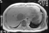
A 63-year-old man was referred to our hospital on April 23, 2003 because of the findings on abdominal CT and a MRI scan that were taken at a local community hospital. The findings showed a single liver tumor measuring 6 cm in diameter in the right lobe (Fig. 1). He presented with a two-month history of intermittent fever that ranged from 37.5 to 38.0℃ and a weight loss of 4 kg (64 kg to 60 kg). The physical examination was unremarkable.
Laboratory investigations revealed that the hemoglobin level was 9.3 g/dL, and the white blood cell count was 8,680/µL with segmental neutrophilia (78.2%). There was no eosinophilia (1.4%). The erythrocyte sedimentation rate was elevated to 97 mm/h (normal range : 0-15 mm/h) and the serum C-reactive protein level was also elevated to 8.18 mg/dL (normal range : 0-0.8 mg/dL). The liver function tests were within normal limits as were the tests for serum alpha-fetoprotein (αFP), carcinoembryonic antigen (CEA) and carbohydrate antigen (CA) 19-9. Serology for hepatitis A, B, or C was negative. No organisms were identified on the cultures of peripheral blood. Intradermal skin tests for Clonorchis sinensis and Paragonimus westermani were negative.
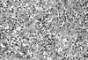
Percutaneous ultrasound guided core biopsy was performed to allow a histological diagnosis of the liver mass, and it confirmed IPT of the liver (Fig. 2). Cultures of biopsed tissue were not done.
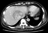
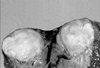
The patient was initially treated with antibiotics (cefpiramide, amikacin, metronidazole, teicoplanin and, imipenem) for 4 weeks and a nonsteroidal anti-inflammatory drug (Naproxen) was prescribed for 8 weeks. Yet the fever continued to spike to 38.0℃ and the lesion still remained on the follow-up CT scan that was done July 13, 2003 (Fig. 3). Therefore extended right hepatectomy was carried out on July 16, 2003, and a tumor, 6 cm in diameter, was found in Couinaud's segment VIII of the liver (Fig. 4). The histologic findings were consistent with the features of IPT (Fig. 5). There were no complications and the fever subsided during the postoperative period. The patient was discharged on the 14th postoperative day, and he is now doing well without any recurrent fever during the follow-up period of 12 months.
DISCUSSION
The exact pathogenesis for IPT of the liver has not yet been well characterized, but the inflammatory pathological pattern and the systemic symptoms including fever and malaise suggested there was an underlying infectious agent. However, in many reports, no causative microorganisms have been identified in the blood cultures, the same as was noted in our case.7
The differential diagnosis from malignant tumor can be difficult, because the radiologic findings of IPT are rather nonspecific.8 Percutaneous biopsy is most reliable method, and it enables us to avoid unnecessary exploratory laparotomy or a hepatectomy when there is an uncertain diagnosis. On rare occasion, an occult adenocarcinoma could be mistaken as an IPT.9 Pseudotumor should be kept in mind in the differential diagnosis of malignant liver lesions.
Numerous studies have shown that the natural history of IPT is one of disease regression. Once the diagnosis of IPT has been confirmed with biopsy, patients with IPT can simply be observed and regular follow-up is performed until the condition resolves itself, or the patient can be medically treated with antibiotics, anti-inflammatory drugs and steroid.10
Although surgical resection is generally regarded as excessive treatment, surgery should be considered in the following situations. The first case is that the systemic symptoms with fever do not resolve in spite of conservative therapy, as was the situation in our case.11 The second situation is that IPT is noted to grow, with or without symptoms, on serial examinations and the imaging studies.12 The last situation is IPT that involves the hepatic hilum, and thic can causes biliary obstruction and portal hypertension.13
Although symptomatic patients may benefit from steroid administration, we selected liver resection as the treatment of choice instead of steroid therapy in this case because there are no clinical reports showing that steroid therapy promotes resolution of IPT.10 For respectable IPTs that are unresponsive to antibiotics, surgery could be a better option rather than steroid therapy or simple observation.

 XML Download