Abstract
Pharyngoesophageal perforation from an exploding bottle is an extremely rare injury. To date, twenty-four cases have been documented in English literature. In this study, we reported two additional cases of pharyngoesophageal perforation by a bottle exploding in the mouth. Explosion of the bottle occurred when the patients removed the cap of a home-made wine bottle with their teeth, which resulted in pharyngoesophageal perforation. The patients were managed by conservative treatment and operative repair, respectively. Both patients had an uneventful recovery. Possible mechanisms and preventive measures are discussed in this study, along with a review of the literature.
The explosion of a drink bottle is a very rare cause of traumatic pharyngoesophageal perforation. Pharyngoesophageal perforation may occur when one removes a bottle cap with their teeth. The rapid pressure increase in the closed oral cavity not only causes laceration of the oral and pharyngoesophageal soft tissues, but also cervical emphysema, pneumothorax, and pneumomediastinum. Pharyngoesophageal perforation by an exploding bottle was first reported by Bowsher et al.1 in 1982. Subsequently, twenty-three cases have been documented in English literature.1-12 This study reports two additional cases of pharyngoesphageal perforation by an exploding bottle, and a review of the literature pertaining to this condition.
An 83 year-old male was admitted because of bleeding from the mouth and dyspnea. He tried to remove the cap of a 1.8-liter plastic bottle of home-made wine with his teeth. The plastic screw cap had blown off and a gas stream burst into his mouth. Neck swelling and dyspnea were observed, even though laceration of the soft palate was repaired before he was transferred to our hospital.
On examination, he was not anemic or shocked. His respiratory rate was 28/min, blood pressure 160/110 mmHg, and pulse rate 128 beats/min. He was not febrile. A flexible larynopharyngoscopy showed laceration and hematoma in the posterior pharyngeal wall and subcutaneous emphysema of the neck. Plain radiographs showed air and widening of the retropharyngeal space, as well as pneumomediastinum. A computed tomography (CT) revealed extensive emphysema of the neck and hematoma obstructing oropharyngeal airway (Fig. 1). He developed acute bleeding from his pharynx and was in respiratory distress. He was intubated immediately and taken to an operating room. A pharyngoscopy showed a perforation in the left side of the posterior pharyngeal wall starting from the inferior part of the nasopharynx and extending to the cricopharyngeus (Fig. 2). The pharyngeal perforation was repaired, but upper esophageal tearing was left unrepaired because of constraints under the pharyngoscopy. Supportive therapy with broad spectrum antibiotics, fluids, oxygen, and a nasogastric tube-feeding was given. Subcutaneous emphysema of the neck gradually improved and pharyngoesophagogram with Gastrograffin showed no leakage 9 days after the operation, so oral feeding was started. He was discharged 14 days after injury, and felt fine at the 1-year follow-up.
A 65-year old female presented with bleeding from the mouth and throat pain. She had been trying to remove the cork of a 750-mL glass bottle of home-made wine with her teeth when it exploded and the cork shot into her mouth. After the explosion she noticed pain in her throat. The glass bottle was not broken and the cork was retrieved from her mouth.
The patient was nervous upon examination, with a temperature of 36.3℃, a respiratory rate of 20/min, a pulse rate of 92 beats/min, and a blood pressure of 170/90 mmHg. Her airway was intact. The posterior pharyngeal wall was lacerated about 2 cm in length and blood clots were seen in the larynx and hypopharynx. Cervical emphysema was noted, but there was no pneumomediastinum or pneumothorax. A lateral neck radiograph and CT showed emphysema in both sides of the neck. Nasogastric tube feeding and prophylactic antibiotics were given because of deep neck infection or mediastinitis. She was feeling well at the 2-year follow-up.
Barotrauma in the aerodigestive tract causes mucosal perforation. Once an abnormally high air pressure is introduced into the potential space of the head and neck, it may extend to deeper structures and to the mediastinum. Through the perforation, normal floras in the aerodigestive tract contaminate the deep space of the head and neck, which may result in deep neck infection or mediastinitis.
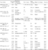
Etiologic agents of barotrauma have been reported as follows: bicycle inner tube,11 tractor tire,13 fire extinguisher,14 and exploding bottle.1 Pharyngoesophageal injury by bottle explosion can develop when patients remove the cap of a bottle with their teeth. Commercially made soft drinks, wine, or beer are safe unless they have been stored under adverse conditions, but home-produced wine and beer are often manufactured in less than ideal circumstances, with considerable build-up of carbon dioxide.7 To date, 24 cases of barotraumatic pharyngoesophageal perforation by an exploding bottle have been reported in English literature and are summarized in Table 1.1-12 The patients were aged 5 to 75 years and consisted of 17 males and 7 females. Most of the cases (22/24) were less than 15 years old, which suggests that educating children and adolescents is essential for prevention.
The pathogenesis of injuries caused by compressed air has been clearly described: the escaping gas enters the mouth and forcibly distends the pharynx and esophagus, causing them to rupture.13 The hurtled cap and broken bottle are also involved in pharyngoesophageal injuries.
Because of its anatomy, the exceedingly friable esophagus is particularly vulnerable at the proximal and distal ends.15 Support for the last 2 inches of the posterior pharynx is somewhat deficient where transverse and oblique fibers of the inferior constrictor muscle converge. This is similarly true in the upper part of the posterior esophagus, where the longitudinal muscle fibers split and leave the mucosal support largely to the circular muscle fibers.13 The distal part of the esophagus has been shown to have segmental defects in the circular muscle layer where the longitudinal muscles end in a conical fashion.13,16 It seems clear that the esophagus at the distal portion is either weaker or less supported by surrounding mediastinal structures.17
When there is a sudden, rapid rise in air pressure in the mouth, the pressure is transmitted to the laryngopharynx and enters the respiratory passages, causing a reflex closure of the glottis. As the cricopharyngeus gives way under the air pressure, it gives rise to a sudden distention of the esophagus.14 The cardia fails to relax as it does in the slower, coordinated, reflex act of swallowing, and the dilated esophagus ruptures.14 Most of the ruptures reported occurred at the lower end of the esophagus (66.6%).18 It appears that the lower end of the esophagus is more vulnerable to bursting from the introduction of pressurized gas.18 If the pressure is slowly released, it may even bring about closure of the cricopharyngeus, creating a closed space between this muscle and the lips, thus causing perforation of the pharynx.9 The posterior wall is the least supported part of the pharynx and is therefore most vulnerable to perforation. In our review, the pharynx was injured in 18 cases,1-7,9-12 the pharynx and esophagus in 5 cases,4,8,11 and the esophagus in 1 case.8 Of 6 cases with esophageal injury, only one had lower esophageal perforation.4 These findings suggest that the explosive pressure of a drink bottle may be insufficient to perforate the lower esophagus.
Pain during swallowing and bleeding from the pharynx are the most common symptoms of perforation. Respiratory compromise may pose an imminent threat to life. In the reported cases, there were 5 cases necessitating endotracheal intubation or a tracheotomy.2-4,11 Subcutaneous emphysema of the neck may be seen on palpation and reflects perforation of the aerodigestive tract. Pneumomediastinum and pneumothorax may occur. Four cases had peumothorax and underwent chest drainage.3-5,9 Fever and leukocytosis suggest complicated infections or abscess formation. Complications of transmural pharyngeal and esophageal injuries include deep neck abscess, mediastinitis, pleural empyema, lung abscess, peritonitis, and sepsis.19 Rubikas reported that overall postoperative mortality in pharyngeal and esophageal injuries was 19%, and the main causes of death were the dissemination of infections and multiple organ insufficiency.19 In the review article of Brinster et al., cervical esophageal perforations were associated with a mortality of 6% (0-16%), whereas thoracic and abdominal perforations were associated with the mortality of 27% (0-44%), and 21% of patients (0-43%), respectively.20 Mediastinitis is one of the fatal complications and is well-known for its high rate of mortality and morbidty. In barotraumatic pharyngoesophageal injury by an exploding bottle, five cases had mediastinitis and one of these cases died of truncal fasciitis and sepsis, postoperatively.2,5,8,12 The overall rate of deep neck infection or mediastinitis was 25% (6/24); these complications occurred in 22% (4/18) of pharyngeal injuries,2,5,6,12 20% (1/5) of pharyngoesophageal injuries,8 and 100% (1/1) of esophageal injuries.8
A physical examination and flexible endoscopy of the oral cavity and laryngopharynx show a laceration on the involved site. Plain radiographs show cervical subcutaneous emphysema, air in the retropharyngeal space, pneumomediastinum, pneumothorax, pleural effusion, and/or pulmonary infiltrates. Gastrograffin swallow defines the site and extent of the perforation and the CT detects the extent of emphysema, infection, and abscess formation.
These patients require immediate attention to the airway, to provide unobstructed breathing and to check for a pneumothorax. Pharyngoesophageal perforation should be repaired as soon as possible, but patients with a small laceration or delay in diagnosis can be strictly observed. Conservative treatment was given in eight patients (33%, 8/24) with no mortality.2,4,7,10,11 If cervical or mediastinal abscess is present, it should be drained externally. Supportive care includes prophylactic antibiotics, nasogastric feeding, and oxygen. Based on a review of literature, most of the patients have a good prognosis if they are properly managed for airway compromise and complications such as mediastinitis.
Suggested measures to prevent injuries from a bottle explosion include the storage of bottles in a cool place, avoidance of jostling or hitting bottles together, directing the cap away from the body or face when opening, storage of bottles on the floor or lowest shelves to reduce the hazards in case the bottle falls or explodes, and avoidance of shaking carbonated drinks.21 School campaigns should be waged because children are the most susceptible. Conspicuous warning labels should be placed on all carbonated drink bottles, and the use of plastic bottles and caps is helpful in reducing incidence of this injury.
Figures and Tables
Fig. 1
Computed tomography of the neck shows extensive emphysema and hematoma obstructing the oropharyngeal airway.

Fig. 2
Pharyngoscopy shows a full-thickness laceration along the posterior pharyngeal wall extending to the cricopharyngeus.

Table 1
Summary of Reported Cases of Barotraumatic Pharyngoesophageal Injuries by Exploding Bottle in the Literature

T, tonsil; OT, oral tongue; SP, soft palate; ICA, internal carotid artery; TP, tonsillar pilla; PPA, parapharyngeal abscess; M, mediastinitis; P, pharynx; UE, Upper esophagus; LE, lower esophagus; CD, cervical drainage; PPW, posterior pharyngeal wall; MD, mediastinal drainage; RPA, retropharyngeal abscess; CE, cervical esophagus; TE, thoracic esophagus; MA, mediastinal abscess; TF, truncal fasciitis; S, sepsis; OP, oropharynx; BOT, base of tongue.
References
1. Bowsher WG, Kenyon GS. Accidental oropharyngeal injury. Br Med J. 1982. 284:1752.
2. Irwin BC. Accidental oropharyngeal injury. Br Med J. 1982. 285:63.
3. Du Plessis HJ, Becker JH. Exploding bottle tops. Br J Surg. 1986. 73:863.
4. Conlan AA, Wessels A, Hammond CA, Beale PG, Katz G. Pharyngoesophageal barotrauma in children: a report of six cases. J Thorac Cardiovasc Surg. 1984. 88:452–453.
5. Wood DJ, Milford D. Accidental pharyngeal rupture from explosion of a bottle into the mouth. Injury. 1986. 17:40.
6. Forer M, Flynn P, Hughes CF, Szasz J. Pneumatic rupture of the posterior pharyngeal wall. Aust N Z J Surg. 1986. 56:89–91.
7. Vallis MP, Gibbin KP. The exploding bottle top and oropharyngeal injuries. Br J Surg. 1986. 73:221.
8. Meyerovitch J, Ben Ami T, Rozenman J, Barzilay Z. Pneumatic rupture of the esophagus caused by carbonated drinks. Pediatr Radiol. 1988. 18:468–470.
9. Bar-Maor JA, Hayari L. Pneumatic perforation of the esophagus in children. J Pediatr Surg. 1992. 27:1532–1533.
10. Efrati Y, Sarfaty S, Klin B, Eshel G, Segal S, Vinograd I. Oral blast injury caused by an accident. Ann Otol Rhinol Laryngol. 1993. 102:528–530.
11. Kraus M, Peiser J, Bartal N, Fliss DM. Cervical subcutaneous emphysema due to oropharyngeal barotrauma. J Oral Maxillofac Surg. 1995. 53:1215–1217.
12. Tostevin PM, Hollis LJ, Bailey CM. Pharyngeal trauma in children--accidental and otherwise. J Laryngol Otol. 1995. 109:1168–1175.
13. Buntain WL, Lynn HB. Traumatic pneumatic disruption of the esophagus. J Thorac Cardiovasc Surg. 1972. 63:553–560.
14. Cole DS, Burcher SK. Accidental pneumatic rupture of oesophagus and stomach. Lancet. 1961. 1:24–25.
15. Bernatz PE. Management of esophageal perforations. Mayo Clin Proc. 1956. 31:671–678.
16. Mackler SA. Spontaneous rupture of the esophagus; an experimental and clinical study. Surg Gynecol Obstet. 1952. 95:345–356.
17. Randolph H, Melick DW, Grant AR. Perforation of the esophagus from external trauma or blast injuries. Dis Chest. 1967. 51:121–124.
18. Gelfand ET, Fisk RL, Callaghan JC. Accidental pneumatic rupture of the esophagus. J Thorac Cardiovasc Surg. 1977. 74:142–144.
19. Rubikas R. Pharyngeal and oesophageal injuries. Injury. 2004. 35:371–378.
20. Brinster CJ, Singhal S, Lee L, Marshall MB, Kaiser LR, Kucharczuk JC. Evolving options in the management of esophageal perforation. Ann Thorac Surg. 2004. 77:1475–1483.
21. Bergeson PS, Sehring SA, Callison JR. Pop bottle explosions. JAMA. 1977. 238:1048–1049.


 PDF
PDF ePub
ePub Citation
 Print
Print


 XML Download