

 PDF
PDF ePub
ePub Citation
 Print
Print


INTRODUCTION
Mucins are highly glycosylated glycoproteins found mainly in the mucus secreted into the respiratory, gastrointestinal and genitourinary tracts.1 They are expressed in various epithelial cells to form the glycocalyx at mucosal surfaces, and act as a lubricant as well as a barrier, protecting cells from dehydration, proteolysis, and infection. Furthermore, they can act as an anti-adhesion molecule,2 serve as a receptor and its cognate binding protein,3 and contribute to signal transduction.4
The currently known mucin species can be divided into two groups, dependent on their structural aspects and biosynthetic routes. Membrane-bound mucins (MUC1, MUC3, MUC4, MUC12) exhibit hydrophobic sequences or "transmembrane domains" responsible for anchoring them in the lipid bilayer and have C-terminal peptides that enter the cytosol. The secretory mucins (MUC2, MUC5AC, MUC5B, MUC6) with one exception (MUC7) possess one or several von Willebrand factor-like D domains, cystein-rich peptides, which function in the oligomerization of mucin monomers and in packaging into secretory vesicles.5 Mucin1 was the gene first discovered in the mucin family,6 which includes 12 different members (from mucin1 to mucin12).7
Mucins in the female reproductive tract have been focused. The epithelial expression of mucin1 in the female reproductive tract and its state of glycosylation are likely to be involved in the transit of oocytes and spermatozoa, and in the uterine implantation of embryos.8 But little is known about mucins in the male genital tract, except for some reports concerning the epithelium of the prostate,9 the epididymis and the finding of mucin8 antigenicity on spermatozoa.10
In this study, we sought to characterize the expression pattern of mucin genes in the human testis, and to evaluate the relationship between the expression of mucin genes and impaired spermatogenesis in the human testis.
MATERIALS AND METHODS
Collection and histological evaluation of the human testis
Thirty human testis tissues were collected from diagnostic testicular biopsies of 30 azoospermic patients. One part of the tissue underwent histological evaluation, and the other part of the tissue (20-30 mg) was stored with RNAlater solution (Ambion, Austin, TX, USA) at -20℃ until preparation of total RNA. The mean age of the patients was 38.5 years (range 31-45 years).
Clinical background
Of the 30 azoospermic patients, every patient had 46 XY, a normal result of chromosome studies. In the hormonal study results, Sertoli-cell-only syndrome patients (13 cases) had increased FSH levels. The rest of the patients (17 cases) were within the normal range. On physical examination, impaired spermatogenesis patients had decreased testis size (right: 12 ± 2.2 mL, left: 12 ± 2.1 m L).
Preparation of total RNA from the tissue samples
The samples were homogenized and prepared using QIA shredder (Qiagen GmBH, Hilden, Germany). Total RNA was prepared with Qiagen RNeasy Kit (Qiagen) following the manufacturer's instructions. The tissue samples were disrupted and homogenized with the buffer RLT containing mercaptoethanol (10 µL/mL). After centrifugation, the supernatants were carefully transferred to a new microcentrifuge tube and one volume (350 µL or 600 µL) of 70% ethanol was added. The samples were put into RNeasy mini columns, and centrifuged for 15 sec at ≥ 8000 g (≥ 10,000 rpm). After washing and air-drying of the RNeasy columns, total RNAs were eluted with 30-50 µL RNase-free water by centrifugation.
To remove the content of genomic DNA, each sample was subjected to DNase-treatment following the protocol of the DNA-free Kit (Ambion, Austin, TX, USA). DNase I buffer and 1 µL of DNase I (2 units) were added to the RNA samples and then they were incubated at 37℃ for 30 min. The DNase inactivation reagents were applied to the samples for 2 min at room temperature. The Dnase-treated RNA samples were collected from the supernatants after centrifugation at 10,000 g for 2 min. After the treatment with DNase, the amount of RNA was determined by spectrophotometer and then stored at -70℃ until use.
Semiquantitative RT-PCR of mucin genes
The procedure for generating cDNA was done using M-MLV reverse transcriptase (Promega, Madison, WI, USA). The reaction mixture of reverse transcription consisted of RT buffer, 5.5 mM MgCl2, 500 µM dNTPs, 2.5 µM oligo-dT(16), 0.4/µL RNase inhibitor and 1.25 U/µL of RTase. The RT reaction mixture (16 µL) and purified RNA (4 µL) were incubated at 25℃ for 10 min and 48℃ for 30 min. RTase was denatured by incubation for 5 min at 95℃.
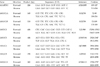
RT product (2 µL) was used directly for the PCR along with specific primers for human mucin genes (Table 1). Complementary DNA of the tissues were amplified by the following PCR conditions: 20 µL of reaction mixture, which consisted of 10 mM Tris-HCl, 1.5 mM MgCl2, 50 mM KCl, and 0.01% gelatin, 200 µM each dGTP, dATP, dTTP and dCTP, 100 pmol of the oligonucleotide primer and 1 unit of Taq polymerase (Promega, Madison, WI, USA). The following amplification profile was used in a Stratagene RoboCycler 96 (Stratagene, La Jolla, CA, USA): 1 cycle of 94℃ for 1 min; 30 cycles of 94℃ for 30 sec, 58-64℃ for 30 sec and 68℃ for 2 min; 1 cycle of 68℃ for 10 min; and a final hold at 4 . After PCR, the 10 µL of PCR products were run on 2% agarose gels containing 0.5 µg/mL of ethidium bromide. The relative amount of mucin mRNAs was calculated according to the ratio of glyceraldehydes-3-phosphate dehydrogenase (GAPDH) bands as an internal control on the agarose gels. The ratio was determined by densitometry after correction for the intensity of the control band using an image analyzer (Vilber Lourmat, France).
Primer design for alternative splicing of human MUC1 gene
For the detection of mucin1 gene, the following primer pair was used: 5'-CCTTTCTTCCTGCTGCTG-3' (forward) and 5'-TGGGCACTGAACTTCTG-3' (reverse). These primers span intron 1 and an alternative splice site of 27 bp at the 5'-end of exon 2 (variants A and B), characteristically leading to two different RT-PCR products of 118 and 145 bp in length.11,12 The two splice variants closely resembled one another. The only difference was the use of two alternative splice acceptor sites for exon 2, located 27 bp apart.
RESULTS
Histological diagnosis of the human testis
In the histological evaluations, 13 samples showed normal spermatogenesis, and these patients were diagnosed as having obstructive azoospermia. The histological findings of the other 17 samples were spermatocyte arrest (4 cases) and Sertoli-cell-only syndrome (13 cases). These patients were diagnosed as having nonobstructive azoospermia. The following study was performed using both the samples with normal spermatogenesis (n = 13) and impaired spermatogenesis (n = 17).
Mucin gene expression in the human testis
The RT-PCR products of human GAPDH (226 bp) as an internal control were detected from all human testis samples. The expression of mucin1, 9 and 13 were found, but RT-PCR products of mucin2, 3 and 4 were not detected in the human testis. Alternative splicing variants of mucin1A (145 bp) and B (118 bp), which were expected by primer design, were detected in the human testis. The RT-PCR products of mucin1, 9 and 13 were confirmed by the direct sequencing (data not shown).
Mucin gene expression in the human testis with normal or impaired spermatogenesis
The RT-PCR products of mucin1, 9 and 13 were found in human testis. In the testis with impaired spermatogenesis, RT-PCR product of mucin1A and B was not found. There was no difference in mucin9 and 13 expression between the testis with normal and impaired spermatogenesis. These results are summarized in Table 2.
DISCUSSION
This study was performed in order to investigate the expression patterns of mucin genes in human testis. In human testis, the mRNA of mucin1, 9 and 13 were detected but the mRNA of mucin2, 3 and 4 were not found. Two types of alternatively spliced mucin1A and B transcripts were expressed in the human testis with normal spermatogenesis. However, there was no expression of mucin1 in the human testis with impaired spermatogenesis. Glycosylated MUC1 proteins would enlarge the space between Sertoli cells and expressing germ cells, interfere with physiological cell functions and probably accelerate the detachment and transit of delayed or abnormal germ cells.12,13 MUC1 expressions were only found in the testis with normal spermatogenesis, not in the spermatocyte. MUC1 isoforms expressed in human spermatids contribute to the glycocalyx of the sperm surface.14 To our knowledge, this study is the first that has detected mucin9 and 13 in the human testis. Mucins may have an important role in spermatogenesis and gamete transport in the human testis.
Relatively little is known about mucin gene expression in the mammalian male reproductive tract, including in humans. HE6, a gene encoding a protein with multiple membrane-spanning domains and mucin-like regions, was detected in the epididymis,15 and MUC8 was found on human spermatozoa.10 Recently, mucin1 expression was confirmed by immunohistochemistry and RT-PCR in human testis with normal spermatogenesis.12 The results further suggested that aberrant expression of mucin1 related to impaired spermatogenesis in human testis. Our study showed that two types of alternatively spliced mucin1 transcripts were detected only in the human testis with normal spermatogenesis. This finding is similar to the previous report.12 Unlike Franke's study, many of our patients were in the late stage of maturation arrest or Sertoli-cell-only syndrome, so we could not observe MUC1 expression in patients in the impaired spermatogenesis group.12
Mucin9 is known to be a precursor to the oviduct specific glycoprotein, oviductin.16,17 As a unique component of oviduct fluid, it participates in fertilization, facilitates early embryonic development, and protects the tubal epithelium.18,19 Hendrix et al.20 reported unexpected identification of mucin9 as an estrogen-dependent product of the adult rabbit endocervix. In this study, MUC9 was first detected in the male reproductive tracts of mouse and human testis. It is an extraordinary result, and the function of MUC9 should be investigated in the male reproductive tracts.
Recently, Williams et al.21 reported that the human mucin13 gene encodes an epithelial and hematopoietic transmembrane mucin. Mucin13 is the orthologue of a murine glycoprotein initially called 114/A10, or mouse cell surface antigen, and it was initially identified by expression cloning using an antibody produced against a murine bone marrow-derived multipotential cell line.22 Mucin13, like mucin1, may play an important role in epithelial responses to damage and infection and in non-malignant epithelial diseases in which barrier function and/or mucin secretion are important, such as inflammatory bowel diseases, cystic fibrosis (CF), and chronic respiratory diseases. The testis has a blood barrier and mucin13 may have a role in the environment of spermatogenesis.
Conclusively, mucin1, 9 and 13 were expressed in the human testis, and they may play a role in spermatogenesis and gamete transport. Further studies are needed on mucin protein expression and localization by immunohistochemistry to evaluate whether mucin1 expression is essential for normal spermatogenesis in human testis.

 XML Download