

 PDF
PDF ePub
ePub Citation
 Print
Print


In recent years, the biological safety evaluation of biomaterials and medical devices has become more globally standardized, concurrent with the publication of the ISO 10993 standard for biomaterial and medical device testing.1 Although an in vitro cytotoxicity test using a mammalian cell culture has been adopted for primary safety evaluation prior to in vivo testing in every national and international standard, the recommended methodologies and cell lines used vary, and accordingly cytotoxicity quoted results from each sample vary according to the standards.2-6
Cytotoxicity tests are recommended for all medical devices as they allow a rapid evaluation, employ standard protocols, produce quantitative and comparable data, and due to their sensitivity, allow toxic materials to be discarded prior to animal testing.6,7 Generally, three types of cytotoxicity tests are used: the extract dilution method, the direct contact method, and the indirect contact (agar diffusion) method.7 The direct contact method enables weak cytotoxicity to be detected because of its high sensitivity,8,9 whereas the extract dilution method is more commonly adopted for the in vitro cytotoxicity evaluation of materials and devices used in the body, since it can be applied to a wide variety of raw materials and finished products that may release toxins from exposed surfaces.10,11 Extraction conditions (time and temperature) are dependent on the physicochemical characteristics of the material being tested and the extraction vehicle.12,13
Recommended conditions may be applied according to the device characteristics and the specific conditions of use.11 These conditions are as follows: a) not less than 24 h at 37℃, b) 72 h at 50℃, c) 24 h at 70℃, and d) 1 h at 121℃.11 Extraction conditions should simulate as closely as possible the conditions under which the device will normally be used. Established cell lines are preferred and can be obtained from recognized repositories. Where a specific sensitivity is required, primary cell cultures and cell lines obtained directly from living tissues should only be used if reproducibility and accuracy of the response can be demonstrated.14-16 However, several problems, such as the preparation of test materials (in the case of the extract method), choice of cell type, the test procedure and method used to quantify results, etc., have been encountered in vitro cytotoxicity tests.17-19 In this study, the cytotoxicity of latex gloves to cultured L-929 cells was determined under various extraction conditions by using the extract dilution method. Since 1980, the cytotoxicity of latex urinary catheters has been widely reported on in clinical and experimental situations using various cell lines, such as, human urothelial cells, V79 cells and L-929 cells.20,21 Therefore, latex gloves are suitable test samples for evaluating the optimal extraction conditions for cytotoxicity tests.
All reagents used for cytotoxicity testing were purchased from Sigma (St. Louis, MO., USA), unless otherwise specified. The powder free latex gloves were purchased from Woo Jin ACT Inc. Seoul, Korea. The gloves were minced and then sterilized using ethylene oxide gas prior to testing.
NCTC clone 929 (L-929, mouse subcutaneous connective tissue) cells were purchased from the American Type Culture Collection (Rockville, MD, USA). The cells were initially cultured and routinely maintained in Dulbecco's modified Eagle's medium (GIBCO BRL, Grand Island, NY, USA) supplemented with 10% fetal bovine serum (FBS; GIBCO BRL) and 1% penicillin-streptomycin (GIBCO BRL) at 37℃ and 5% CO2 in a humid environment. The collected L-929 cells were then plated in 24-well microculture plates at a density of 2×10⁵ cells/well in complete culture medium.
According to the extraction time and temperature, the extraction conditions used for extracting latex gloves were as follows: 1) 24 h at 37℃; 2) 72 h at 37℃; 3) 72 h at 50℃. In addition, the solvents used for extracting the sample were distilled water (DW), 9 g/L sodium chloride (saline) in DW, and culture media with or without 10% heat-inactivated FBS. The extracts were 2-fold serially diluted by adding fresh culture media (2×) containing 10% FBS. After the L-929 cells had been seeded at a density of 2.0×10⁵ cells/well into a 24-well plate in duplicate and incubated at 37℃ for 24 h, the medium was replaced with the diluted extracts and then the cells were incubated for a further 24 h.
After incubation, each well was washed with phosphate-buffered saline (PBS) and stained with 0.2% crystal violet (CV) solution dissolved in 2% ethanol for 20 min. The stained cells were lysed with 0.5% sodium dodecyl sulfate solution in 50% ethanol and then transferred to a 96-well plate. The absorbance of each well was measured at 610 nm using an automatic microplate reader (Spectra Max 340, Molecular Device Inc., Sunnyvale, CA, USA). The cytotoxic potential of the latex gloves to the cultured L-929 cells was expressed in terms of the relative viability. Relative cell viability was defined as the percentage of the optical density of a diluted extract containing medium to the optical density of the corresponding non-treated control.
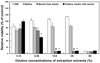
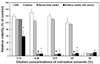
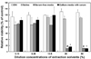
In this study, the extract dilution method was used to determine optimal extraction conditions required to the cytotoxicity testing of latex gloves to cultured L-929 cells, with respect to time, temperature, and solvent composition. When the latex gloves were extracted in either DW or saline at 37℃ for 24 h, no cytotoxicity was observed at a 25% concentration or less (Fig. 1). In contrast, extracts obtained using media with or without serum resulted in marked toxicity even at 12.5% of the initial concentration. These results showed that extraction by culture medium provided a more sensitive evaluation of glove cytotoxicity. Similarly, when extraction was done for 72 h at 37℃, neither DW nor saline resulted in noticeable cytotoxicity at low extract concentrations (≤25%, Fig. 2), though a slight cytotoxicity was observed, but only a concentration of 50% for both showed distinct cytotoxicity. Extraction in culture medium with or without serum at 37℃ for 72 h produced results similar to observed for 24 h at 37℃. Moreover, cytotoxicity was observed for the latex gloves even at a concentration of 6.25%. It was found that the culture medium with serum was slightly more sensitive for cytotoxicity evaluation than that without serum. Regardless of the extraction solvents, the extraction condition 72 h at 50℃ was less sensitive, than the above-mentioned conditions. As shown in Fig. 3, cytotoxicity of the latex gloves, extracted in DW or saline, to L-929 cells was not occurred at all the extract concentrations. In the cases of the extraction in the culture medium with or without serum, the cells were non-viable due to the latex glove cytotoxicity at higher extract concentrations (25% and 50%).
Generally direct contact methods have various advantages, because they mimic physiological conditions, the zone of diffusion represents a concentration gradient of toxic chemicals, and require no extraction preparation. But the major difficulty of this assay is the risk of physical trauma to cultured cells from either sample movement or crushing due to sample weight.7,22 In vitro methods of cytotoxicity testing should quantify cell viability and growth, and be correlated with in vivo methods or animal tests.11,23 Although an extract dilution test provides a quantitative comparison with positive and negative controls, it presents difficulties in terms of preparing sample extracts. When culture medium is used as the extraction solvent, both polar and non polar components are extracted from the sample, and as was found in the present study, a culture medium containing serum had higher toxicity than normal saline.24 It has also been documented that the toxicities of polymeric biomaterials, would seem to be overwhelmingly due to their leachables.25
As shown in Figs. 1, 2 and 3, the results of cytotoxicity tests using the extract dilution method showed dose-dependent relationships and produced diverse results. Unlike extractions at 37℃ for 24 h or 72 h, extractions for 72 h at 50℃ were insufficient to evaluate sample cytotoxicity. These results seemed to be due to the degradation species released from the latex that might induce cytotoxic effects.
In conclusion, the use of culture medium with or without serum led much higher cytotoxicities than extractions performed with DW or saline. Additionally, extractions at 37℃ for 24 h or 72 h were found to be more sensitive and effective at evaluating latex glove cytotoxicity by the extract dilution method. Our results suggest that a combination of two or more extraction methods compatible with the physicochemical characteristics of test materials is required for in vitro cytotoxicity testing.
 XML Download