

 PDF
PDF ePub
ePub Citation
 Print
Print


INTRODUCTION
Schwannomas are common tumors of the spine, but extremely rare in the upper cervical region. Spinal schwannomas are typically benign tumors that arise from schwann cells of nerve roots, and compromise 29% of all spinal tumors.1 They usually develop inside the dural sac, but 10-15% of schwannomas extend along the spinal nerve root inside the foramen and usually present with extradural and sometimes extraspinal components.2 Although some spinal schwannomas have been reported to invade the vertebral body2,3 and cause osteolytic lesions, schwannomas of the C-1 and C-2 roots seldom invade the lateral mass. This is because they can extend through the wide space behind the lateral mass and also there is a lack of intervertebral foramen at the C-1 vertebra. This report documents a case of schwannoma of the C-1 root that invaded the transverse process and articular facet of C1, and resembled an exophytic bone tumor.
CASE REPORT
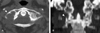
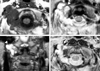
A 60-year-old woman presented to the clinic complaining of neck pain for one and a half months without any neurological abnormality. The pre-operative computed tomography (CT) scan revealed extensive erosion with a marginal sclerosis or destructive pattern involving the right transverse process and articular facet of C-1 (Fig. 1). Axial and coronal magnetic resonance imaging (MRI) scans revealed a multilobulated mass that aggressively invaded the lateral mass of C-1 and had moderate enhancement with the indefinite tumor margin (Fig. 2). Although a benign schwannoma was the most likely diagnosis, we could not exclude the possibility of a malignant tumor, including a malignant nerve sheath tumor or a primary malignant bone tumor.
A right, extreme lateral approach to the lesion was used to access it. After reflection of the skin and muscle layers, the C-1 and C-2 vertebrae and the margin of the foramen magnum were exposed. The tumor was firm and yellowish in color. After C-1 laminectomy was performed on the right side, we could observe the transverse and articular processes of C-1, which were severely eroded by the tumor's mass. The tumor's capsule was opened and internal decompression was performed. The tumor was attached to the first cervical nerve medially and was subtotally removed due to adhesion with the vertebral artery. The surgical opening was closed in layers according to established protocols.
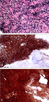
The histopathological diagnosis was a benign schwannoma composed of interwoven bundles of spindle-shaped, fibrillated cells (Fig. 3). An immunohistochemical investigation disclosed a strong reactivity for S-100 protein and vimentin. The patient experienced no neurological deficit or any complication following surgery and she was discharged from the hospital seven days later. Follow-up MRI scans showed good stability of the atlanto-axial joint, but revealed remnants of the tumor which required the patient to undergo radiotherapy (Fig. 2).
DISCUSSION
Schwannomas represent 29% of spinal tumors, although neurofibromas are encountered less frequently. Schwannomas are typically solitary, circumscribed and encapsulated tumors eccentrically located on proximal nerves or the spinal nerve roots.1 Although schwannomas are relatively common benign tumors in the spinal region, there are only a few reports of C-1 neurinomas including schwannomas.2,4,5 In 1988, Guidetti and Spallone5 reported three cases of C-1 tumors and Lot and George2 reported four cases of C1 neurinomas (three neurofibromas, one schwannoma) in 1997.
Schwannomas are tumors that gradually increase in size, and only occasionally are they accompanied by pressure erosion of the adjacent bone, resulting in a concave deformity of the bony surface or an enlargement of the canal. Therefore, extensive erosion of the bone is usually considered uncommon for benign schwannomas, but common for malignant bone tumors. Schwannomas can involve bone through three possible mechanisms. First, a tumor can arise centrally within the bone (intraosseous schwannoma). Second, an extraosseous tumor can cause secondary erosion of the bone by pressure. Lastly, an extraosseous tumor can also arise within the nutrient canal and grow in a dumbbell-shaped configuration, resulting in the enlargement of the canal.6 In this case, the surgical findings indicated that the lesions were of the extraosseous type and were considered to originate in a C-1 nerve root. C1-root schwannomas can extend through wide spaces behind the lateral mass without extensive invasion because there is no intervertertebral foramen for the C-1 nerve root. Therefore, we speculate that the third mechanism, rather than the second mechanism, is related to the tumor growing in our patient and lead to the extensive expansion into the lateral vertebral mass.
In an article on expansile vertebral bone lesions, extradural schwannomas were not included because osteoblastomas, aneurismal bone cysts, and giant-cell tumors are among the tumors usually considered when a patient has an expansile spinal lesion.7 Inoaka et al.6 presented two unusual cases of benign extraosseous neurinomas associated with aggressive intravertebral extension in the thoracic and lumbar regions. The case reported here is an extremely rare example of a C-1 schwannoma with invasion of the lateral vertebral mass.
 XML Download