

 PDF
PDF ePub
ePub Citation
 Print
Print


INTRODUCTION
The syndrome arthrogryposis multiplex congenita (AMC) is characterized by the presence of multiple contractures in several body areas present at birth.1 AMC occurs in approximately 1 in 3,000 births,2 and the principal cause of arthrogryposis is decreased fetal movements (fetal akinesia).1 Amyoplasia congenita is the most common type of arthrogryposis, in which the extremities are usually involved symmetrically, which occurs in 1 in 10,000 live births. Here the author describes a case of amyoplasia congenita involving only the lower extremities in a premature baby.
CASE REPORT
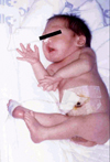
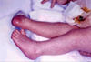
The patient was the first male baby of healthy, non-consanguineous parents; the mother is a 28-year-old preschool teacher, and the father is a 33-year-old businessman. There was no history of medication use or radiation exposure during pregnancy. The mother was a smoker prior to the pregnancy. However, a history of decreased fetal movement was present, and the intrauterine presentation was breech. The child was delivered at a private hospital by Cesarean section at 35+4 weeks due to the breech presentation and premature membrane rupture (9 hours). The Apgar score was 7 and 8 at 1 and 5 minutes, respectively. The birth weight was 1,970 g (within ± 2 SD), the head circumference was 31 cm (within ± 2 SD), and the length was 47 cm (within ± 2 SD). He was transferred from the hospital immediately after birth because of the abnormal leg features. His heart rate was 148 per minute, and the respiratory rate was 52 breaths per minute at time of admission. His activity was moderate to poor, but he was not floppy. Also, his inhalation power was poor. A physical examination upon admission demonstrated the following cylindrical contractures involving the lower extremities (Fig. 1, 2): (1) knees were fixed symmetrically in the hyperextended position with a range of motion (ROM) of 0°; (2) hips were fixed in the flexed position by nearly 90° and limited in abduction with a ROM of 0-45°, and (3) thin subcutaneous tissues and absence of skin creases of the lower extremities. The sensation of the lower extremities was intact, although the deep tendon reflexes could not be evaluated due to the fixed extension of both knees. The feet, upper extremities, and shoulders showed no deformities. His hands lacked Simian lines. Otherwise, he had no remarkable physical findings, except for an undescended right testicle.
Results from laboratory studies were normal, including serum creatine kinase, blood culture, TORCH, newborn screenings, ultrasonography of the brain, abdomen and hip joints, and electromyography. The chromosome analysis revealed a normal 46 XY karyotype. Simple knee X-rays showed increased soft tissue densities in the joint cavities.
He underwent physiotherapy beginning 7 days after birth. From 12 days of age, his activity showed marked improvement and began eating well. He was discharged at 49 days of age. At discharge, his body weight was 3,550 g, with a feeding volume of 80 ml per bottle. His activity was also good. He demonstrated improved ROM, 0-5° of the knee joints, and 0-60° in the abduction of the hip joints.
DISCUSSION
AMC is a descriptive term (arthro=joint, gryp=curved, multiplex=multiple, congenita=present at birth). Arthrogryposis is a congenital disorder characterized by multiple rigid joint deformities,3 and these contractures are usually non-progressive.1 This condition may be isolated, or it may be associated with other types of congenital abnormalities as part of a larger syndrome, possibly affecting the central nervous system.1 The exact pathogenesis of arthrogryposis is currently unknown, but each type includes decreased fetal movements (fetal akinesia), and there is a direct correlation between early onset of fetal akinesia and severity of contractures at birth.1 Typically, joint development is normal at first, except for the lack of fetal movement, and the formation of extra connective tissues around the joints.4 This fixes the joints and aggravates the contractures.4 Although the degree of joint contractures and associated clinical abnormalities varies from patient to patient, there are five common clinical characteristics: 1) affected extremities are fusiform or cylindrical with thin subcutaneous tissue and lack skin creases; 2) rigidity of joints; 3) dislocation of joints, especially the hips and the knees; 4) atrophy and even the absence of muscles or muscle groups, and 5) intact sensation, although the deep tendon reflexes may occasionally be diminished or absent.3 The above deformities are usually symmetric, and the severity increases distally in the affected limb.5
The arthrogryposis can be classified into three main groups: Group 1 affects only the limbs; Group 2 affects the limbs, and either the trunk, craniofacies, or viscera; and Group 3 affects the limbs and involves central nervous system dysfunction.1
Group 1 disorders, "amyoplasia" (A=no, myo=muscle, and plasia=growth), are the most common type of arthrogryposis seen clinically.1 It has an incidence of 1/10,000 live births and comprises approximately one-third of arthrogryposis cases.1 The limb findings in amyoplasia congenita are usually symmetric, mostly involving all four extremities. However, in some patients only the lower extremities are affected, and more rarely only the upper extremities are affected, while the trunk is spared.1 Patients with AMC affecting only the upper extremities show typical positioning, in which the shoulders are internally rotated, sloped, and rounded. The arms are extended and the wrists and hands are flexed, creating the so-called "police-man tip" position.2 When the lower limbs are affected, nearly all cases involve contractures around the hips, most of which are severe.6 Flexion, abduction and external rotation contractures are most common in the hip joints, and dislocation occurs in approximately one-third of patients.6 The knees may be flexed or hyperextended, and the feet are often in an equinovarus position.2 The muscle mass of the limbs is diminished and replaced by fibrous tissue, giving the extremities a slender appearance.1,2
In approximately 10% of individuals with amyoplasia, abdominal structural anomalies with congenital hernia are accompanied by renal abnormalities or abdominal wall defects.7 Facial features are not pathognomic, though a round face with micrognathia and a small upturned nose are often observed.2 A midline nevus flammeus of the forehead is present in 75% of patients.8 Other malformations reported in patients with amyoplasia include: cryptorchism, hydrocele, absence of the vagina/uterus, and tacheoesophageal fistula.5 Fetal intrauterine presentation is breech in nearly one-third of patients with amyoplasia.8 Amyoplasia congenita is sporadic in occurrence, and one study reported no recurrences in families of 135 affected patients.9
The preservation of normal sensation in amyoplasia suggests that the lesion site is located in the anterior horn cells, with predominance of specific neuronal segments.10 The pattern of neurosegmental involvement makes it highly unlikely that muscles or peripheral nerves are the sites of the primary pathological lesion.10 A study found that the most common form of arthrogryposis showed a reduction in the number of spinal cord anterior horn cells. During the developmental period, the anterior horn cells are highly susceptible to insults, like hypotension. Thus, it was postulated that hypotension in the developing fetal spinal cord may occur in this form of disorder. In several previous studies, all dissections of the spinal cord of patients with neurogenic arthrogryposis demonstrated an absence or decreased number of anterior horn cells in the cervical, thoracic, and lumbar segments.11
It is still not known whether the spinal cord (anterior horn cell defect)10,11 or the muscles are primarily affected in amyoplasia. In a study by Hall et al.,8 histological examination of muscle showed non-specific changes, such as the replacement of muscle with fibrofatty scar tissue. Also, the observed muscle spindles were normal. An electromyographic study revealed both neuropathic and myopathic changes at different muscle sites, even within the same patient.8 Based on these findings, muscle biopsy is generally not recommended to establish the diagnosis.
The prognosis of patients with amyoplasia congenita is fairly optimistic. By age 5, 85% of children are ambulatory and completely independent with regard to daily activities.6 Primary goals of management for these conditions are ambulation and maximum upper limb mobilization.2 Aggressive physical and occupational therapy beginning soon after birth is crucial to attaining optimal physical function.2 Educationally, most children will function in a regular classroom setting at the appropriate grade level, with only 4% in special programs.6
In this case, the patient's mother had a history of fetal akinesia during pregnancy, and he was delivered by Cesarean section due to breech presentation. Severe, fixed hyperextended contractures of hip joints and fixed flexed contractures of knee joints were noted along with thin subcutaneous tissue and a lack of skin creases at birth, though sensation was intact in the lower extremities.
Therefore, the author reports a case of amyoplasia congenita involving only the lower extremities with sporadic occurrence in a premature baby.
 XML Download