

 PDF
PDF ePub
ePub Citation
 Print
Print


INTRODUCTION
Obesity is an independent risk factor for cardiovascular disease.1,2 In particular, abdominal visceral adipose tissue is believed to have harmful effects on vascular endothelial function through various interactions of coagulants and inflammatory molecules.3 The vascular endothelium serves as an endocrine organ, secreting several vasoactive substances, such as nitric oxide. These substances prevent the development of atheroma. Obesity is independently associated with vascular endothelial dysfunction (VED,) and VED is a key event in the early development of atherosclerosis. Therefore, VED may be an important link between obesity and atherosclerosis. Weight reduction has been reported to improve VED.4 However, because obesity is frequently accompanied by other cardiovascular risk factors and metabolic diseases, the ability of reduced visceral adiposity alone to improve VED in obese patients has been difficult to determine.
A simple, non-invasive pulse wave analysis base technique was recently used to evaluate vasomotor endothelial function.5,6 After demonstrating that albuterol,7 a β2 -agonist, reduces wave reflection by activating the L-arginine-NO pathway, many studies have shown that an assessment of global endothelial function can be performed by examining changes in the peripheral waveform in response to inhalation of β2-agonists. Changes can be measured by tonometry and quantified according to the augmentation index (AIx).5,6 The present study utilized the advantages of pulse wave analysis, i.e., its simplicity, repeatability, operator-independency and non-invasiveness. We evaluated the impact of substantial weight reduction on VED in premenopausal obese women without complications or any other cardiac risk factors to determine whether weight reduction alone was associated with improved VED.
MATERIALS AND METHODS
Subjects
The same healthy, premenopausal obese women included in a previous study8 were enrolled in the present study. These subjects were visiting a university hospital obesity clinic and were 20 to 45 years-old (mean age: 33.81 ± 7.13) with a body mass index (BMI) ≥ 25 kg/m2. Obesity was defined according to the guidelines established by the 'Redefining Obesity and its Treatment'- a Joint Enterprise by the Regional Office for the Western Pacific of the World Health Organization and the International Association for the Study of Obesity and International Obesity Task Force. Subjects with any of the following co-morbidities were excluded: hypertension (systolic blood pressure > 140 mmHg or diastolic blood pressure > 90 mm Hg), dyslipidemia {LDL (low density lipoprotein) cholesterol ≥ 160 mg/dL, HDL (high-density lipoprotein) cholesterol < 40 mg/dL, or triglyceride ≥ 200 mg/dL}, type 2 diabetes, coronary heart disease, stroke, thyroid disease, and depression. Information was obtained by administering a self-report questionnaire to patients and by clinical examination and blood tests. Current smokers and those who had undergone a commercial diet program or received anti-obesity medication during the previous three months were also excluded. As in our previous study, obese subjects were matched with healthy, premenopausal non-obese women for clinical, metabolic, and lipid characteristics. Initially, 42 obese patients were enrolled, but six dropped out over the 12-week study period, leaving a total of 36 subjects. All subjects were given informed consent, and the study protocol was approved by the Institutional Review Board of Ewha Womans University, Mokdong Hospital.
Data collection
All examinations were performed during the follicular phase of the menstrual cycle, within 10 days of the onset of menstruation. Subjects received dietary and exercise consultations every two weeks for three months, during which BP, height, weight, and waist and hip circumferences were measured.
Anthropometric measurements
Body weights were measured to an accuracy of 0.1 kg, with subjects wearing light clothing. Height was measured to an accuracy of 0.1 cm. The waist circumference was measured at the mid-point between the upper iliac and lower costal level at the end of normal expiration, to an accuracy of 0.1 cm, and the hip circumference was defined as the largest measurement around the hip area. Body mass index (BMI) was calculated as the weight in kilograms divided by the square of the height in meters (kg/m2).
Body fat measurements
Percent body fat (%) was measured using bioelectrical impedance analysis while subjects were in a fasting state (Inbody 2.0, Biospace Co., Seoul, Korea), and body fat distribution was determined by computed tomography (GE High Speed Advantage CT scanner, GE Co., Waukesha, USA). Single slices of abdominal subcutaneous adipose tissue and visceral adipose tissue (measured in cm2) were taken at the 4-5 level of the lumbar spine, as described elsewhere.9 The attenuation interval was -40 to -140 Hounsfield units.
Blood tests
Plasma glucose, total cholesterol, HDL cholesterol, LDL cholesterol, triglyceride, and non-esterified fatty acid (NEFA) concentrations were measured after overnight fasting for at least 12 hours. Plasma insulin was measured by radioimmunoassay. Estradiol levels were measured by microparticle enzyme immunoassay (Axsym, Abbott, Abbott Park, Illinois, USA). Levels of high-density C-reactive protein (hsCRP) were measured by nephelometry using BNII (Dade Behring, Liederbach, Germany) with a minimum detection concentration of 0.0175 mg/dL. Homeostasis Model Assessment (HOMA) scores were calculated to evaluate insulin resistance.10
Evaluation of vascular endothelial function
Pulse wave analysis using the provocative pharmacological test was performed by administering salbutamol or nitroglycerin (NTG) to determine VED.
Pulse wave analysis
A pulse wave is composed of two waves, i.e., an aortic wave produced when the left ventricle contracts and a reflected wave produced when the aortic wave is reflected from peripheral vessels. The velocity of the reflected waves is increased when the arterial walls are stiff. Accordingly, in cases with stiffened arteries, systolic pressure augmentation occurs during the systolic phase. Based on this principle, radial artery pressure was measured using a high-fidelity micromanometer (SphygmoCor, Medical ECONET Co., Sydney, Australia). In practice, the artery is compressed between a sensor and the underlying structures, and, thus, the intra-arterial pressure is transmitted through the arterial wall to the sensor. Data were collected directly using a portable computer. After 20 sequential waveforms had been acquired, integral software was used to generate an averaged peripheral waveform and a corresponding central waveform. Further analysis was conducted to determine AIx and the ascending aortic pressure, as described previously.7,11,12 AIx was defined as the difference between the first (P1) and second (P2) peaks of the central arterial waveform, expressed as a percentage of pulse pressure.13,14 In this study, repeated pulse wave analyses at one-week intervals in ten non-obese volunteers showed a coefficient of variance (CV) of 1.4 ± 2.3% for AIx.
Measurements of aix after salbutamol or ntg administration
AIx was evaluated after administering nitroglycerin or salbutamol, which induce endothelial-independent vasodilation and endothelial-dependent vasodilation, respectively. AIx was determined during the peak response which occurred 3 minutes after administering 0.6 mg of nitroglycerin. The difference in AIx before and after this administration was measured and is depicted as ΔAIx-NTG. Twenty-five minutes after administering nitroglycerin, 2.5 mg of nebulized salbutamol (Ventolin® nebules), which acts on vascular endothelial cells through the L-arginine-NO pathway, was administered by inhalation using a mask. Thiry minutes later, the pulse wave analysis was again carried out to measure AIx. The difference between AIx before and after salbutamol administration was then determined and is depicted as ΔAIx-Salb.5,7,11 The dosage and timing of the administration of these drugs were based on pilot studies and previous experience. Pilot studies confirmed that 15 minutes was sufficient to allow NTG-induced hemodynamic changes to return to baseline, but that a longer period was required for salbutamol. Therefore, NTG was always administered first, and followed by salbutamol 25 minutes later.
Diet and exercise program
For effective weight control, a diet and exercise program was utilized, and subjects were educated regarding living habits. To maintain a low calorie/low fat diet, subjects received 1,000 kcal per day during the program. Initially, subjects exercised for half an hour a day, 3 times a week. The length and frequency of exercise were then increased every other week, reaching 40-60 minutes a day and 4-5 times a week. Subject compliance was evaluated at every visit by examining the dietary diary and weight loss log. Subjects were administered 20 mg of fluoxetine daily, as well as a thermogenic agent (pseudoephedrine 60 mg, theolan 100 mg) as a supplementary measure. To avoid side-effects, drugs were administered for the first 4 weeks only. Three months later, physical examinations, obesity measurements, and pulse wave analysis were repeated.
Statistical analysis
All data are expressed as means±SD. Changes in ΔAIx-NTG, ΔAIx-Salb and all other parameters versus weight reduction were analyzed using a paired t-test. To identify parameters affecting (AIx-Salb, a multiple linear regression model was constructed using the step-wise method, after adjusting for BMI with (AIx-Salb as the dependent variable. hsCRP was log transformed to achieve a normal distribution. The level of significance was set at p < 0.05 (two-tailed). Statistical calculations were performed using the SAS system for Windows (version 8.0, SAS Inc. Raleigh, NC, USA).
RESULTS
Anthropometric and clinico-metabolic responses to weight reduction
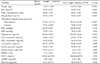
After 12 weeks of the diet and exercise program, all subjects were re-evaluated. The average weight reduction was 7.96 ± 3.47 kg (11% of base-line), with losses of 5.29±2.59 kg (13% of baseline) in fat mass (p < 0.001) and a marked reduction in BMI and waist circumferences. The visceral and subcutaneous fat areas were reduced by 21.88 ± 20.39 cm2 and 49.01 ± 53.76 cm2, respectively (p < 0.001). Diastolic pressure was slightly reduced, without a significant change in systolic pressure. The levels of total cholesterol, LDL cholesterol, and triglycerides were all reduced after weight reduction, but the serum HDL cholesterol level did not show an increase following weight reduction. Fasting glucose, fasting insulin, HOMA scores, and hsCRP levels were lower following weight reduction, while estradiol levels increased (Table 1).
Vascular reactivity and hemodynamic changes by weight reduction. Baseline heart rate, mean arterial pressure (MAP), and baseline AIx were not significantly changed by weight reduction. Changes in heart rate and MAP provoked by the administration of NTG or salbutamol before weight loss were similar to changes after weight reduction (Table 2). Pulse wave analysis combined with provocative pharmacological testing demonstrated preserved endothelium-independent vasodilation in healthy premenopausal obese women (ΔAIx-NTG: 31.36 ± 9.80% before weight reduction vs. 28.25 ± 11.21% after weight reduction, p > 0.05) and an improvement in endothelial-dependent vasodilation following weight reduction (ΔAIx-Salb: 10.03 ± 6.49% before weight reduction vs. 19.33 ± 9.28% after reduction, p < 0.001) (Table 2).
Relationship between anthropometric and clinico-metabolic parameters and weight reduction
Improvements in endothelial-dependent vascular function following weight reduction (increase in ΔAIx-Salb by weight reduction) were related to reductions in visceral adipose tissue area (r= -0.62, p < 0.001), percent body fat (r= -0.35, p < 0.05), BMI (r= -0.33, p < 0.05), fasting insulin (r= -0.38, p < 0.05), and HOMA scores (r= -0.375, p < 0.05). Moreover, step-wise multiple regression analysis identified visceral adipose tissue area (β= -0.57, p < 0.001) as the most significant independent parameter accounting for improved endothelial-dependent vascular function following weight reduction (Table 3).
DISCUSSION
Obesity is characterized by VED, and the degree of VED is predicted by body fat distribution, independent of metabolic and other hemodynamic parameters.15,16 This association between obesity and VED, an early feature of cardiovascular disease, is of great importance because it may provide clinicians with new preventive strategies against atherosclerosis in obese patients. Recently, a prospective study demonstrated VED improvement following substantial weight reduction in postmenopausal obese women with no additional risk factors.4 However, the study tested VED by determining hemodynamic and rheologic responses to L-arginine, a technique that may be inadequate for screening or clinical applications. A simple and noninvasive form of pulse wave analysis to determine VED was recently introduced.5,6 In the present study, we used pulse wave analysis, combined with provocative pharmacological testing, to determine VED. To our knowledge, this is the first report to prospectively test the impact of weight reduction per se on VED by pulse wave analysis. The main finding of this study is that substantial weight reduction over a 12-week period can improve VED, and that visceral fat reduction is most strongly related to VED improvement in premenopausal obese women.
Relationship between visceral adiposity and VED
Adipose tissue acts as an endocrine organ and appears to secrete metabolic pro-inflammatory molecules. These cytokines are known to contribute to vascular inflammation and to regulate vascular endothelial function, an early indicator of atherosclerosis development. Moreover, visceral fat appears to produce these cytokines more actively than other adipose tissues. Visceral fat cells show higher rates of catecholamine-induced lipolysis and express higher numbers of β1- and β2-adrenergic receptors and levels of glucocorticoid receptor mRNA, thus explaining the importance of fat distribution.17 Aging and estrogen deficiency can play a role in changes in vascular endothelium. After menopause, the possibility of obesity-related complications is likely to increase. Other cardiovascular risk factors, such as hypertension, diabetes mellitus, hypercholesterolemia, and smoking are also associated with VED. Thus, we confined our study to premenopausal obese women without additional cardiovascular risk factors in order to reduce the effect of such factors on VED.18 In a previous study, we found that provocative pharmacological testing using NTG showed preserved endothelium-independent vasodilation in premenopausal, healthy, obese women. However, changes in AIx after the introduction of salbutamol nebulas, which induces vasodilation through vascular endothelial cells, was less evident in the obese group than in the non-obese group. The results of the present study suggest that endothelial vasodilatory function is reduced in premenopausal healthy obese women despite a similar level of arterial stiffness, thus confirming that visceral fat per se is highly associated with VED.
Relationship between reduced visceral adiposity and improved VED
Vascular endothelial function is affected by cardiovascular risk factors, hormones, and medications. Factors that affect VED include hypertension,19 hypercholesterolemia,20 aging,21 estrogen,22 smoking, diabetes mellitus,23 cholesterol-lowering agents,24,25 hormone replacement therapy,26,27 and physical inactivity.28 Weight reduction improves many of the conditions frequently associated with or co-occur with obesity. This raises the question of whether the restoration of vascular endothelial function by weight reduction in obese people is due to visceral adiposity reduction, an improvement in cardiovascular risk factors, or increased physical activity. A recent study4 demonstrated that a reduction of visceral adipose tissue in obese women is a more important factor than increased physical activity or improvement of cardiovascular risk factors in improving VED. The study4 showed that a reduction in adipose tissue by liposuction surgery improved VED and highlighted the importance of visceral adiposity reduction in and of itself in improving VED in obese women.
Pulse wave analysis for the evaluation of VED
Vascular endothelial cell function was measured using pulse wave analysis. The results of pulse wave analysis following the administration of salbutamol or NTG has been shown to correlate with the results of invasive methods based on acetylcholine infusion.5 The augmentation index was used to assess VED in the present study. Compared to established methods for assessing vascular endothelial function, AIx combined with a drug challenge is a non-invasive, simple, and convenient method that is useful for the early detection and assessment of VED.5,29 Recently, however, some questions have been raised about the validity and clinical utility of pulse wave analysis. Wide margins of error were found when comparing centrally measured and peripherally derived AIx.30 AIx was also found to be dependent on heart rate and blood pressure during IV infusion of beta-adrenergic drugs, such as isoproterenol.31 However, the inhalation of albuterol was not accompanied by significant alterations in heart rate and MAP.5 We also confirmed in a previous study that a substantial change in heart rate or MAP was not induced by NTG or salbutamol. In the present study, changes in ΔAIx-Salb by weight reduction were not related to a small change in heart rate or diastolic or systolic pressure (Table 3). Moreover, as the absolute value of AIx might not be of importance in follow-up studies,32 we used AIx with provocative pharmacological testing in this study. Since all subjects were premenopausal women free of cardiovascular disease, it remains to be determined whether our findings can be applied to men, children, postmenopausal women, and cardiovascular patients.
In conclusion, we have demonstrated that a reduction in visceral adiposity is significantly related to improvements in VED, as determined by radial artery pulse wave analysis, following weight reduction. In this context, clinicians may choose to focus on the reduction of visceral adiposity, as much as on the control of other risk factors, for the prevention of atherosclerosis in obese women.


 XML Download