

 PDF
PDF ePub
ePub Citation
 Print
Print


INTRODUCTION
Dieulafoy's lesion is an uncommon disease entity that causes gastrointestinal tract hemorrhage. It was first described by Gallard in 18841 and later named after Dieulafoy in 1898.2 Dieulafoy's vascular malformation may cause massive, potentially life-threatening and often recurrent gastrointestinal bleeding.3 Although most of the Dieulafoy's lesions occur in stomach within 6 cm of the gastroesophageal junction,4 these lesions have also been reported in the duodenum, jejunum and colon.5 Matuchansky described two cases of jejunal bleeding from the rupture of a solitary large submucosal artery.6 We report here on a case of jejunal Dieulafoy's lesion that was identified by computed tomography (CT) and enteroclysis.
CASE REPORT
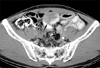
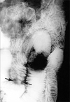
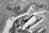
A 35-year-old man presented with a 1-month history of dizziness, palpitation and indigestion. He had a feeling of fullness in his epigastrium and he had esperienced a weight loss (5 Kg/month). On physical examination, the patient was pale with a blood pressure of 130/70 mmHg. The admission laboratory tests revealed a Hb of 9.1 g/dL, a Hct of 30%, normal platelets and normal coagulation parameters. The stool was positive for blood, but there is no bleeding focus in esophagogastroduodenoscopy, and colonoscopy. A CT scan was performed for further evaluation, and it showed a mass in the lumen of the jejunum with subtle contrast enhancement. Minimal bowel dilation was also seen proximal to the jejunal mass (Fig. 1). Enteroclysis revealed a short segment of jejunal stricture with central ulceration (Fig. 2). The angiogram did not show any extravasation or staining. Segmental resection of the jejunum was then done. There was a jejunal mass with stricture and adhesion, and the surgical pathology revealed a large caliber persistent submucosal artery protruding through a mucosal defect (Fig. 3).
DISCUSSION
Dieulafoy's lesion is characterized by massive gastrointestinal hemorrhage and hypotension. Histologically, it is a large caliber persistent artery that is usually 1-3 mm in diameter, and it courses tortuously through the submucosa and comes in intimate contact with the mucosa.7 The pathogenesis and bleeding mechanism are not well understood. It may involve aging that may lead to elongation and tortuosity of a submucosal artery.8 Chronic gastritis, alcohol and anti-inflammatory drugs have been suggested as a trigger for the acute bleeding in this gastric lesion. Others have suggested that the close proximity of the pulsating vessel to the epithelium lining may mechanically damage the overlying mucosa and induce ulceration. Dysplastic changes are evidenced by the subintimal fibrosis and the loss of elastic fibers adjacent to the necrotic artery wall: the thinning or loss of circular fibers of artery may predispose it to sudden hemorrhage.7 There have been several additional reports of jejunal Dieulafoy's lesion.9-11 However, to the best of our knowledge, there has been no well documented radiological report. Therefore, we report here the radiological findings of Dieulafoy's lesion with a literature review. In our case, the gross specimen revealed an ulcerative lesion with hematoma that was thought to be a jejunal mass on the CT scan. The serosal surface of the lesion was congested and adhered with fibrosis. This finding could explain the stricture of the jejuneum on the enteroclysis.
In conclusion, Dieulafoy's lesion could be one of differential diagnoses of the small bowel mass or stricture, and especially in young patients who suffer from massive, recurrent gastrointestinal bleeding.
 XML Download