

 PDF
PDF ePub
ePub Citation
 Print
Print


INTRODUCTION
Pelvic organ prolapse (POP) is a condition in which the vagina, bladder, rectum or some digestive organs are prolapsed from their normal anatomical locations in the pelvis. The prolapse is due to the damage of the intrapelvic supporting tissues. In the United States about 400,000 surgeries are performed yearly to treat pelvic organ prolapse.1 Luber et al. (2001) report that pelvic organ prolapse is found in 1.7 per 1,000 women in their 30s. This figure significantly increases with age as 18.6 per 1,000 women in their 70s have pelvic organ prolapse.2 The prevalence rate of pelvic organ prolapse then is directly proportional to age. Hence, it becomes important for physicians to recognize pelvic organ prolapse as a major quality of life issue in aging women.
In patients with pelvic organ prolapse, the prolapsed pelvic organ affects neighboring organs such as the bladder and urethra. Subsequently, this can lead to lower urinary tract symptoms such as frequent urination, dysuria, urinary incontinence, urgency, hesitancy, etc.3 Richardson et al. (1983) reported that the kinking of the urethra that occurs in pelvic organ prolapse induces urethral closure, and an increase in the urethral closure pressure.4 Bump et al. (1988) and Rosenzweig et al. (1992) reported an increase in the urethral closure pressure in urodynamic studies of pelvic organ prolapse patients.5,6
In patients with pelvic organ prolapse, the increase in urethral closure pressure, due to the pelvic organ prolapse, results in the improvement of urinary incontinence. However, if the prolapse is surgically corrected, there may be a worsening of the incontinence.6 Thus, when performing a surgery to correct the pelvic organ prolapse, it is necessary to anticipate how it will affect postoperative urethral functions. Currently, there are no studies or reports on how pelvic organ prolapse affects changes in urethral function, or what factors are related to those changes.
This study performed manual reduction specifically on patients with anterior vaginal wall prolapse, and preoperatively estimated the urethral function changes through maximum urethral closure pressure (MUCP) and functional urethral length (FUL). Then, the urethral function changes for pelvic organ prolapse were predicted postoperatively by studying the differences of those changes by age and stage.
MATERIALS & METHODS
This study included 139 female patients with anterior vaginal wall prolapse who came to the YUMC between March 1999 and May 2003. All patients had a urethral pressure profile, and underwent a manual reduction of the prolapse. The patients' anterior vaginal wall defects were then diagnosed as Aa or Ba according to the POP-Q (quantification) system to evaluate pelvic organ prolapse.7
This study was approved by the hospital's institutional review board. We compared and analyzed the patients' characteristics in age, pelvic organ prolapse stage, delivery history, pregnancy history, body mass index (BMI), concomitance of stress urinary incontinence (SUI), and urethral pressure profile. The urethral pressure profile was estimated using a 10 French catheter. The SUI in the subjects was diagnosed according to the definition of the International Continence Society.8
Statistical analysis was conducted through analysis of variation (ANOVA), chi square testing, and t-test using SPSS software windows version 10.0 (SPSS INC, Chicago, IL, USA) at a 5% level of significance (p < 0.05). Pearson correlation coefficient was used to verify the relationship between urethral pressure profile and BMI, pregnancy history, and delivery history, as well as the relationship between pelvic organ prolapse stage and SUI.
RESULTS
We investigated the age, stage, pregnancy history, delivery history, and BMI of 130 subjects with pelvic organ prolapse whose urethral pressure profiles were estimated after preoperative manual reduction. First, the patients' ages comprised the following: 3 (2.2%) cases occurred in those under age 30, 13 cases (9.4%) in the 40s, 53 cases (38.1%) in the 50s, 54 cases (38.8%) in the 60s, and 16 cases (11.5%) in those over age 70. The stages of the subjects consisted of 3 (2.2%) cases at stage I, 35 cases (25.2%) at stage II, 76 cases (54.7%) at stage III, and 25 cases (18.0%) at stage IV. The statistical analysis excluded very small groups (such as the 3 cases under age 30, and the 3 cases at stage I) for more accurate statistical analysis (Table 1).
The characteristics of the subjects were as follows: mean BMI 23.47 ± 2.94 Kg/m2, mean pregnancy history 5.75 ± 2.55 times, mean delivery history 3.66 ± 1.55 times, mean maximum urethral closure pressure (MUCP) 69.22 ± 23.08 cmH2O, and mean functional urethral length 36.67 ± 8.93 mm (Table 2).
The MUCP did not vary much with age as it was 71.69 ± 26.36 cmH2O in the 40s, 73.64 ± 21.31 cmH2O in the 50s, 66.09 ± 22.47 cmH2O in the 60s, and 57.75 ± 23.55 cmH2O in the 70s and over (p=0.07). On the other hand, the functional urethral length was significantly shorter in women in their 50s and 60s than in women in their 40s. This is based on the estimation that the FUL was 43.23 ± 7.43 mm in the 40s, 36.63 ± 8.71 mm in the 50s, 34.66 ± 7.74 mm in the 60s, and 39.06 ± 12.00 mm in those age 70 and over (p=0.01) (Table 3).
The MUCP according to the stage of the pelvic organ prolapse was not significantly different. The MUCP was 68.74 ± 20.31 cmH2O at stage II, 67.01 ± 20.22 cmH2O at stage III, and 79.00 ± 30.85 cm H2O at stage IV (p=0.07). The functional urethral length at stage IV was much longer than that at stage III (34.92 ± 7.71 mm at stage III, and 40.32 ± 11.00 mm at stage IV, while 37.66 ± 9.42 mm at stage II) (p=0.02). There was no significant difference between stages II and III (Table 4).
We investigated the correlation between the urethral pressure profile and the BMI, pregnancy history, and delivery history. The correlation coefficients between the patients' BMI, MUCP, and FUL were 0.08 and 0.05 respectively. This means that the BMI did not affect the urethral pressure profile. The correlation coefficients of the pregnancy history, MUCP, and FUL were -0.12 and -0.18 each. This suggests that the pregnancy history did not affect the urethral pressure profile as well. The delivery history also did not influence the MUCP and the functional urethral length as the Pearson correlation coefficients were -0.04 and 0.02.
In addition, of the 139 patients diagnosed as having pelvic organ prolapse, 83 (59.71%) patients reported symptoms of SUI. The SUI concomitance by the patients' ages was found to be in 2 (66.7%) patients under age 30, 10 (76.9%) patients in their 40s, 26 (49.1%) patients in their 50s, 36 (66.7%) patients in their 60s, and 9 (56.25%) patients age 70 and over. The patients' age and SUI prevalence rate were not related based on the results obtained through chi square testing (p=0.5) (Table 5).
The SUI concomitance by pelvic organ prolapse stage was shown in 2 (66.7%) cases at stage I, 26 (74.29%) cases at stage II, 47 (62.64%) cases at stage III, and 8 (32%) cases at stage IV. Using these results in a chi-square test demonstrated a negative relationship between the pelvic organ prolapse stage and the SUI prevalence rate.
DISCUSSION
Many patients coming to the department of obstetrics and gynecology show the pelvic organ prolapse manifestation. It is especially evident in elderly women as the prevalence rate for pelvic organ prolapse is directly proportional to age. Hence, it is important that physicians inquire into quality-of-life issues that may be a direct consequence of the prolapse.
Pelvic organ prolapse occurs due to complicated disorders of muscles, nerves, and connective tissue that cause pelvic supporting tissue weakness. This kind of pelvic support weakness simultaneously induces the urethral hypermobility known as stress urinary incontinence.9 Snooks et al. (1985) reported that this incontinence is associated with damage to pudendal nerves distributed in the muscles around the urogenital diaphragm, external anal sphincter, and perineum. Busacchi et al. (1999) also reported the relationship between pudendal nerves and pelvic organ prolapse.10,11 Moreover, the anatomical prolapse of intrapelvic organs caused by the weakness of the pelvic supporting structures affects the neighboring organs such as the bladder and urethra. Hence, prolapse has been reported to cause symptoms such as frequency, dysuria, incontinence, urgency, and hesitancy. It also investigates kinking of the urethra to induce mechanical urethral occlusion. This ultimately results in increased urethral closure pressure.3,4
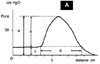
Thus, there is a close relationship between pelvic organ prolapse and stress urinary incontinence. However, the lower urinary tract symptoms caused by pelvic organ prolapse or stress urinary incontinence are nonspecific and subjective. Urodynamic studies are then used to objectively evaluate lower urinary tract dysfunctions that may be caused by pelvic organ prolapse or stress urinary incontinence.12 In a urodynamic study, the urethral functions can be estimated through a urethral pressure profile which includes the MUCP and FUL. The urethral closure pressure (Pclose) can then be calculated by taking the difference between urethral pressure (Pure) and vesicular pressure (Pves): (Pclose=Pure-Pves). The functional urethral length can be figured out using the length of the urethral pressure change. This is determined by doubling the length of a section from a vesicular pressure point to the maximum urethral closure pressure 13 (Fig. 1).
The MUCP has been reported to be increased by urethral mechanical closure due to the urethral kinking found in patients with pelvic organ prolapse.14-17 However, patients with stress urinary incontinence have been reported to show a decrease in the MUCP which leads to a decrease in functional urethral length.16,18,19 Subsequently, the incontinence symptoms are improved in pelvic organ prolapse patients because of increases in the MUCP and functional urethral length (owing to the prolapse). However, the incontinence can be more pronounced after the the prolapse is corrected.14
The understanding of the correlation between pelvic organ prolapse and stress urinary incontinence is essential in predicting and ameliorating the onset of stress urinary incontinence in the postoperative period. Accordingly, this study attempted to predict the postoperative urethral function changes by examining the urethral pressure profile, and evaluating urethral functions both pre- and postoperatively.
To maximize the goals of the study, the stage of the pelvic organ prolapse was corrected using the stage of the anterior vaginal wall prolapse. Aa or Ba was then used to correspond to the dependent portions of the anterior vaginal wall defect (as per the POP-Q system).
However, the changes in the pelvic organ prolapse stage and the anterior vaginal wall prolapse stage were only shown in a total of 2 cases, with most of the patients having the anterior vaginal wall defects in the dependent portion.
In the current study, the patient's age did not affect the onset of stress urinary incontinence (Table 5). However, the presence of a higher stage of prolapse decreased the onset of the stress urinary incontinence (Table 6). This result harmonizes with reports that the maximum urethral closure pressure and functional urethral length are increased in pelvic organ prolapse patients.14-17 Nevertheless, this result is contrary to the report of Chaikin et al. (2000). This study found that severe pelvic organ prolapse accompanies stress urinary incontinence more frequently.21 Therefore, further investigation evaluating pelvic supporting muscle weakness, and urodynamic studies is warranted.
In this study, the MUCP and FUL (obtained through the urethral pressure profile after the correction of the pelvic organ prolapse with preoperative manual reduction) were statistically identified regardless of the patient's age or the stage of the pelvic organ prolapse. The effects of age and stage on the urethral function are caused by the mechanical occlusion of the urethra (due to the anatomical dislocation of the pelvic organ), and are temporary. Davic et al. (1983) performed the correction of the pelvic organ prolapse through a pessary or manual reduction, and reported that the corrected MUCP was lower than the initial one in the urethral pressure profile.17 Although the results of Davic et al. may also represent the temporary phenomenon of urethral occlusion, it does not necessarily mean that the urethral function changes are due to the prolapse. Therefore, the preoperative and postoperative urethral functions of the patient with pelvic organ prolapse are thought to be identical regardless of the patient's age or prolapse stage. Thus, the possible postoperative stress urinary incontinence and its related lower urinary tract symptoms are more a manifestation of mechanical occlusion rather that prolapse.
Consequently, the stress urinary incontinence that is absent in pelvic organ prolapse patients can be produced after corrective surgery. Therefore, implementation and comparison of urodynamic studies before and after pelvic organ prolapse correction can be useful in determining whether it is necessary to provide additional surgery or treatment for preventing SUI.20,21
In this study, 83 (59.71%) pelvic organ prolapse patients experienced accompanying stress urinary incontinence. However, urethral occlusion was implicated for the decrease in the maximum urethral closure pressure. If additional investigation is done into the urethral pressure profile before correcting pelvic organ prolapse through manual reduction, it is possible to evaluate the direct consequences of the prolapse on urethral functions.


 XML Download