

 PDF
PDF ePub
ePub Citation
 Print
Print


INTRODUCTION
Orbital wall fractures are a common result of orbital injury. An orbital wall fracture may occur when the orbit is bluntly struck by an object with dimensions greater than the anterior orbital rim. Fractures of the orbital wall may lead to enophthalmos, limitation of ocular motility, diplopia, and altered sensation in the distribution area of the infraorbital nerve.1,2 Many surgeons immediately repair orbital floor fractures based on their demonstration by orbital computed tomography (CT). Other surgeons follow the post trauma course for development of vision-disabling diplopia or facial asymmetry. The advent of CT in the late 1970s and early 1980s produced a fairly uniform protocol that remains in widespread use: large fractures portending enophthalmos can be repaired within 2 weeks of the injury; small fractures are repaired if clinically significant diplopia does not resolve within 2 weeks of observation.3-6 The causes of ocular motility disturbance and diplopia are: extrusion of the extraocular muscles or orbital soft tissues, injury in the extraocular muscles, edema or hemorrhage in the fat tissue within the orbit, and vertical deviation of the eyeball.7 In short, ocular motility disturbance has two principal causes: functional disability of muscles due to the paralysis of the extraocular muscles and limitation caused by nearby structures. However, it is difficult to identify the exact cause of the ocular motility disturbance. Although there are many studies on orbital fracture management, few studies have been published comparing the preoperative clinical characteristics, including CT findings, and persistent postoperative ocular motility disturbance. In the case of ocular motility disturbance and diplopia due to orbital fracture, studies are required to identify their causes after proper treatment for orbital wall fracture. By performing CT and various ocular motility tests before and after the surgery, this study analyzes the functions of the extraocular muscles and determines factors affecting postoperative ocular motility disturbances.
MATERIALS AND METHODS
Between February 2001 and January 2003, 45 eyes of 45 patients with orbital wall fractures, whose medical records could be traced back at least 6 months, underwent surgical repair in our hospital. Records were reviewed to confirm the presence of an orbital fracture, to identify demographic information, and to determine the cause, location, degree and type of each fracture. Extraocular motility limitation, field of binocular single vision and a Hess screen test were performed. All patients underwent a coronal and axial CT to determine the presence of fracture, the involvement of the orbital wall, the location of the orbital wall, the degree of fracture, and the degree of muscle or soft tissue entrapment.
Orbital computed tomography analysis
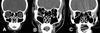
The involvement of orbital walls was classified into medial, inferior, medial and inferior, or inferior and lateral. The location of the orbital wall was classified as anterior, middle, or posterior. This was according to the location of the section containing the largest fracture from the 1.0 mm thickness image of CT scanning (Fig. 1, 2).
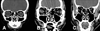
The degree of fracture was classified into mild (minimal orbital expansion and soft tissue herniation), moderate (moderate expansion and prolapse), or severe (significant expansion and prolapse), according to the degree of orbital volume expansion and soft tissue prolapse (Fig. 3).8
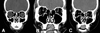
The degree of muscle or soft tissue entrapment was classified as free (not continuous with any bony density in any of the scan), hooked (continuous with bone along one side, but with the majority of the muscle continuous with only soft tissue or fat densities), and entrapped (adjacent to bone, nasally and temporally) (Fig. 4).8
Ocular motility test
Ocular motility tests are conducted after trauma, 1 day after the surgery, and 6 months after the surgery. The ocular motility limitation (ranging from 0 to 4) is measured in mm based on the degree of limitation of ocular motility of the injured eye, compared with the normal eye in conjugate gaze. The sum of the ocular motility limitation of each 4 directions is graded.
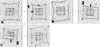
The degree of diplopia was quantified by measuring the field of single binocular vision, ranging from zero to three. Zero corresponds to no diplopia, one to mild (diplopia appears more than 30 degrees from the primary position), two to moderate (diplopia appears more than 30 degrees between 10 and 30 degrees from the primary position), and three to severe (diplopia appears within 10 degrees from the primary position), respectively (Fig. 5).
The extraocular motility disturbance was graded in four cardinal directions by the Hess screen test, zero to three, and added-up. 0 indicates normal, 1 indicates mild reduction of extraocular muscle power, 2 indicates moderate, and 3 indicates severe, compared with the normal Hess screen test results (Fig. 6).
The indication for surgical intervention included enophthalmos more than 2 mm, persistent diplopia, positive sign of forced duction test, and extraocular muscle entrapment. Surgical reduction was performed with a 0.85 or 1.0 mm thickness MEDPOR® barrier sheet (Porex, 15 Dart Road Newnan, GA, USA) implantation, performed through the transconjunctival approach under standard techniques.
SPSS for window Ver. 10.0 was used to compute routine statistics, including Chi-square tests, independent t-tests, and paired t-tests. The significance level used for all statistical tests was 0.05.
RESULTS
The 45 eyes of 45 patients with orbital wall fractures who underwent surgical repair were reviewed. The average age was 24.8 years (ranging from 8 to 52 years). The surgery was performed, on average, 8.5 days (ranging from 4 to 22 days) after trauma.
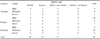
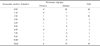
According to the CT result, 15 patients (33%) had a fractured orbital wall in the medial position, 18 patients (40%) in the inferior, 11 patients (24%) in the medial and inferior position, and 1 patient (2%) in the inferior and lateral position (Table 1).
As for the location of the fracture, 22 patients (49%) had a fracture in the middle, 20 patients (44%) in the posterior and 3 patients (6%) in the anterior position (Table 1). As for the degree of fracture, 23 patients (51%) were confirmed to have a moderate fracture, 12 patients (27%) were confirmed to have a mild fracture and 10 patients (22%) suffered from a severe fracture (Table 1).
In the case of the incarceration pattern of extraocular muscles, a hooked pattern appeared in 24 patients (53%), a free pattern in 19 patients (42%), and entrapped in 2 patients (4%) (Table 2).
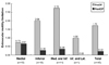
The sum of ocular motility limitation was 1.73±2.46 before the surgery, but showed a significant decrease to 0.22±0.77 6 month after the surgery (Fig. 7, p<0.005).
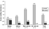
The sum of the degrees of extraocular motility disturbance before the surgery, based on the Hess screen test, was 1.42 ± 1.62, but showed a significant decrease to 0.22 ± 0.70 6 months after the surgery (Fig. 8, p < 0.005).
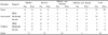
Before the surgery, 33 patients had diplopia and 12 patients experienced no diplopia. There was no statistically significant relation among the existence of diplopia, the fractured orbital wall, the fractured position, the degree of a fracture, and the incarceration pattern. Before the surgery, mild diplopia was seen in 6 patients, moderate diplopia in 17 patients and severe diplopia in 10 patients. However, seven patients successfully recovered from diplopia, and there was significant improvement as to the degree of diplopia after the surgery (Table 3, p < 0.005).
There was no significant difference between the location and degree of fracture and the incarceration pattern of the six patients with moderate or severe diplopia and of the other patients six months after surgery. However, the sum of ocular motility limitation and the degree of extraocular motility restriction in the patients with moderate to severe diplopia showed a significant difference from before surgery to six months after surgery. In the diplopia group (n=6), the sum of ocular motility limitation was 5.67 ± 4.18, and the degree of extraocular motility disturbance was 3.67 ± 2.42 before surgery. In the no diplopia group (n=39), the sum of ocular motility limitation was 1.13 ± 1.38, and the degree of extraocular motility disturbance was 1.08 ± 1.16 (Table 4, 5, p < 0.005).
DISCUSSION
The management of orbital fractures remains controversial. Careful history, physical examination, and CT scans are essential components in the evaluation and subsequent management of patients with orbital wall fractures. Fractures of the orbital wall may arise from hydraulic forces and increased orbital pressure (hydraulic theory), or from direct trauma to the inferior orbital rim, causing buckling of the floor (buckling theory).1,9-11 Symptoms of orbital floor fractures include orbital pain, enophthalmos, hypoesthesia in the V2 distribution (infraorbital: cheek and teeth), and diplopia. Eyelid ecchymosis, subcutaneous emphysema, ptosis, epistaxis, lacrimal system injuries, and pupillary dilation may be associated with orbital floor fractures. Thorough ocular examination is necessary; in particular, special attention is required to assess extraocular motility and forced ductions for extraocular muscle entrapment, ischemia, or hemorrhage. Furthermore, radiographic visualization with CT is essential to detail soft tissue not visible with conventional plain films.12,13
Orbital surgery is not risk free. Potential surgical complications must be considered with the decision to proceed with surgery.14-20 One goal of this study is to predict postoperative ocular motility disturbance, and to correlate CT findings and ocular motility disturbance. Diplopia persists in a significant number of patients following surgical treatment of an orbital wall fracture, even when complete surgical reduction is performed within 2 weeks after the traumatic incident. Diplopia is probably due to a myogenic or neurogenic cause, and, depending upon the degree of deviation, may require surgical correction.21 Lisman et al. has reported that diplopia becomes permanent if a Volkmann's type ischemic contracture of the rectus muscle is left untreated.12 Gillbard et al. noted a relationship between fracture edges and the position of the inferior rectus muscle on coronal CT scans as a sign predicting persistent diplopia.22
According to the studies on the ocular motility disturbance in orbital fracture, Converse et al.22 reported that 80% of orbital fracture patients had an ocular motility disturbance. Greenwood et al.23 reported that 98% of orbital fracture patients had extraocular disability and 89% had diplopia. In addition, Kim and Won24 reported that 100% of orbital fracture patients showed an ocular motility disturbance, 45% of whom suffered from supraduction disturbance, 38% of whom suffered from supraduction and abduction disturbance, and 17% of whom suffered from abduction disturbance. Cha et al.25 showed that 100% of orbital fracture patients showed ocular motility disturbance, 92% of whom suffered from supraduction and infraducion disturbance, 5% of whom suffered from abduction disturbance, and 3% of whom suffered from supraduction and abduction disturbance.
Twenty-six patients (58%) who were subject to our study had ocular motility limitation. Thirty-three patients (73%) had diplopia, 15 patients (46%) had horizontal diplopia, and 18 patients (54%) of them had vertical diplopia. Ocular movement was successfully recovered by surgical reduction performed within 2 weeks after the trauma.
Ocular motility disturbance after the surgery was more related to various ocular motility test results rather than CT findings. Ocular motility disturbance can remain after surgery if ocular motility limitation and extraocular motility disturbance is significant after trauma. Therefore, patients should be given a thorough explanation as to their case, and secondary surgery of extraocular muscles performed.
Additional studies on the various tests to examine functions of extraocular muscles are required to identify and analyze the exact cause of ocular motility disturbance.


 XML Download