

 PDF
PDF ePub
ePub Citation
 Print
Print


INTRODUCTION
Cancer of the cervix is the most common form of cancer for women in developing countries, the second most frequent cancer worldwide, and the most frequent gynecological cancer seen in Korean women.1,2 If cervical carcinoma can be detected early, the prognosis is excellent and complete control may be expected in the premalignant status.3-5 The Pap test, developed by Papanicolaou in 1940s, reduced the incidence and mortality rate of cervical cancer significantly. In the USA, the Pap test reduced the death rate of cervical cancer patients by 70% and it presently saves approximately 10,600 women's lives annually.6 However, reasonable test performance using a competent laboratory results in false negative rates of about 15 - 45% for cervical neoplasia, and supplemental tests that make up for the shortcoming of the cervico- vaginal method are required.6,7
Since Hans Hinselman in Hamburg, Germany, applied colposcopy to cervical cancer in 1925, colposcopy has been used as the other most required test, together with the Pap test, in the detection of cervical cancer and premalignant lesion. The drawbacks of colposcopy, however, are that a large number of patients can not be examined as it requires a specialist with great experience who is excellent in diagnosis. It also requires expensive equipment. It takes a long time for diagnosis. Another shortcoming of colposcopy is the difficulty of its application to general screening and mass screening, as the equipment cannot be moved readily.8,9
Adolf Stafl at the Wisconsin University College of Medicine, USA, developed cervicography in 1981.8 The principle of cervicography is based on colposcopy. However, cervicography obtains objective test materials with the use of a special camera. The procedure is to take pictures of the outside of cervix, develop the pictures and interpret the pictures. Hence, compared with colposcopy, the advantages of cervicography are that it is relatively inexpensive, the equipment can be moved readily and specialists can perform the interpretation objectively and reproducibly.10 Cervicography has been reported to be applicable to mass screening for cervical cancer, and it can facilitate the selection of therapy for patients with abnormal cells detected by the Pap test.11
Recently, regardless of the findings of the Pap test, several institutes have performed cervicography in combination with the Pap test screening. Therefore, we evaluated the effectiveness of applying cervicography in combination with the Pap test for the primary screening of cervical neoplasia.
MATERIALS AND METHODS
Materials
Among all patients screened by cervicography at the Yonsei Cancer Detection Center of the Department of Obstetrics and Gynecology, Yonsei University College of Medicine from January to July 2003, 294 patients who were examined by the Pap test and cervicography as a screening procedure for cervical cancer and taken by the subsequent colposcopy directed biopsy or surgical resection as required, were analyzed. Patients whose results of cervicography had a technical defect were excluded from this study. The mean age of the subjects was 43 years, and the ages ranged from 19 years to 83 years.
Methods
The Pap test was performed first, followed by cervicography. A cytobrush or spatula was used to obtain specimens for the Pap test. The results were interpreted according to the Bethesda classification: negative for intraepithelial lesion or malignancy (N), atypical squamous cells of undetermined significance (ASC-US), atypical squamous cells can not exclude HSIL (ASC-H), low-grade squamous intraepithelial lesion (LSIL), highgrade squamous intraepithelial lesion (HSIL), and squamous cell carcinoma (SCC).
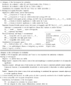
Cervicography was performed with a cerviscope (NTL lab Ltd, Korea), and we used Ektachrome film (ASA 200). The procedure was to insert a speculum, expose the cervix of uterus adequately, remove mucus or blood with cotton swabs, apply 5% acetic acid, and examine the condition of the cervix for 15-20 seconds and note any discharge and bleeding. After applying the acetic acid again, the camera was focused and two cervicograms were taken within 30 seconds. The results of cervicograms were analyzed by the new cervicogram program developed in Korea, and this program was recommended by Korean Woman's Cancer Research Foundation of Catholic Medical Center (Table 1).
To analyze the data, the Pap test classified as atypical squamous cells of undetermined significance (ASC-US), atypical squamous cells can not exclude HSIL (ASC-H), low-grade squamous intraepithelial lesion (LSIL), high-grade squamous intraepithelial lesion (HSIL), and squamous cell carcinoma (SCC) were considered as positive, and suspicious atypia (S1, S2) and positive (PH, PL, and PC) on a cervicogram were considered as positive. The diagnosis of histology above cervical intraepithelial neoplasia (CIN) I was considered as positive. Data was analyzed using parametric and nonparametric statistics, SPSS 10.0 (Chicago, IL, USA). Differences were considered significant when the probability of the error was below 5% (p < 0.05).
RESULTS
The result of screening tests and histology
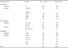
Among 294 women, the Pap test was negative for intraepithelial lesion or malignancy in 130 cases (44.2%), ASC-US in 45 cases (15.3%), ASC-H in 4 cases (1.4%), LSIL in 46 cases (15.6%), HSIL in 46 cases (15.6%), and SCC in 23 cases (7.8%). Cervicographies were negative (N1, N2) in 55 cases (18.7%), benign atypia (B1, B2) in 147 cases (50.0%), suspicious atypia (S1, S2) in 80 cases (27.2%), and positive (PH, PL, PC) in 12 cases (4.1%). The histopathological diagnosis were cervicitis in 131 cases (44.6%), CIN I in 48 cases (16.3%), CIN II in 11 cases (3.7%), CIN III in 25 cases (8.5%), carcinoma in situ of cervix in 31 cases (10.5%), and invasive cervical carcinoma in 48cases (16.3%) (Table 2).
The correlation of screening tests and histology
Among 294 women, the Pap test was negative in 130 cases (44.2%) and positive in 164 cases (55.8%). The sensitivity of the Pap test was 72.0%, the specificity was 64.6%, the positive predictive value was 72.0%, the negative predictive value was 64.6%, the false positive rate was 28.0%, and the false negative rate was 35.4%. The instances of a positive result on the Pap test and negative in histology were 46 cases (15.6 %). The instances of a negative result on the Pap test and positive on the histology were 46 cases (15.6%). The cervicographies were negative in 202 cases (68.7%) and positive in 92 cases (31.3%). The sensitivity of cervicography was 39.6%, the specificity was 79.2%, the positive predictive value was 70.7%, the negative predictive value was 31.0%, the false positive rate was 29.3%, and the false negative rate was 49.0%. The instances of a positive result on cervicography and a negative result on histology were 27 cases (9.2%), and the instance of a negative result on cervicography and a positive result on histology were 99 cases (33.7%). 101 cases (34.4%) were normal by both tests, and 193 cases (65.6%) were positive by either of the two screening tests. When combining the two screening tests, the sensitivity was 79.3%, the specificity was 51.5%, the positive predictive value was 67.4%, the negative predictive value was 66.3%, the false positive rate was 32.6%, and the false negative rate was 33.7%. The instances of a positive result on both screening tests, but with a negative result on histology were 63 cases (21.4%). 34 cases (11.6%) were negative on both tests and positive on histology (Table 3, 5).
The correlation of cervicography based on Pap smear and histology
For 130 women with negative Pap test, 101 cases (77.7%) were negative and 29 cases (22.3%) were positive on cervicography. For patients with negative Pap test, the sensitivity of cervicography was 26.1%, the specificity was 79.8%, the positive predictive value was 41.4%, the negative predictive values was 66.3%, the false positive rate was 58.6%, and the false negative rate was 33.7%. For women with negative Pap test, the instances of a positive result on cervicography and a negative result on histology were 17 cases (13.1%), and the instances of a negative result on cervicography and a positive result on histology were 34 cases (26.2%). For 164 women with positive Pap test, 101 cases (61.6%) were negative and 63 cases (38.4%) were positive on cervicography. For women with positive Pap test, the sensitivity of cervicography was 44.9%, the specificity was 78.3%, the positive predictive value was 84.1%, the negative predictive value was 32.7%, the false positive rate was 15.9%, and the false negative rate was 67.3%. For women with positive Pap test, the instances of a positive result on cervicography and a negative result on histology were 10 cases (6.1%), and the instances of a negative result on cervicography and a positive result on histology were 65 cases (39.6%) (Table 4, 5).
Comparison of diagnostic accuracy according to screening tests
Compared with the Pap test, the diagnostic accuracy of cervicography was inferior because of its lower sensitivity, positive/negative predictive value and higher false positive/negative, even if its specificity was higher. Combining the two screening tests may be partially helpful as the sensitivity of the combined screening tests was higher than the Pap test alone, and together the two tests had a higher negative predictive value and lower false negative rate. Because of its low specificity and low positive predictive value, however, and considering its cost and effectiveness together, the combined tests have limitations as screening tests. We assessed the accuracy of cervicography even in the patients that were classified based on their Pap test results, and although the specificity was slightly higher (79.8%, 78.3% > 64.6%) and the sensitivity was lower (26.1%, 44.9% < 72.0%), the positive/negative predictive value and the false positive/negative rate were not significantly different.
The consistency of cervicography for Pap test was analyzed with Kappa test, and the result was fair (κ=0.71).
DISCUSSION
Cervical cancer is the most common cancer in the reproductive organs of women. Over 450,000 new cases were detected annually, and over 230,000 women die due to this disease with 80% of these deaths occurring in developing countries.1,2 In developed countries, where the screening program has been actively applied, the incidence as well as the mortality rate has been decreased significantly.12-15 The significant decreases in cervical cancer incidence and mortality can be largely attributed to the success of widespread Pap test. The onset and the natural history of cervical cancer are well established. In addition, the early diagnosis of cervical cancer is feasible as its primary lesion is readily accessible. Therefore, cervical cancer is a curable disease if the appropriate treatments are administered at the early stage of disease.
Over the past several decades, numerous studies have been performed to develop screening methods for cervical cancer, and presently, research is actively ongoing to develop methods that will allow doctors to detect premalignant lesions effectively. The Pap test that was developed in 1942 is currently the most frequently and widely used method worldwide as an individual screening test and as a mass-screening test. Nevertheless, its shortcomings of the low sensitivity and the high false-negative results (15-45%) have been reported,7,8 and the main factors contributing to the false-negative rate were specimen collection, laboratory error, and deficiencies in laboratory quality assurance system.16-18 Giles et al. reported that the Pap test failed to detect approximately 30% of invasive cancer and 58% of premalignant lesion of the uterine cervix.19
Hans Hinselman in Hamburg, Germany applied colposcopy to the screening of cervical cancer for the first time in 1925. The advantages of colposcopy are its ability to locate the low infiltrated lesions, to determine the extent of the lesions, and its high accuracy. Colposcopy has been used widely as a diagnostic tool, together with the Pap test, for cervical cancer and premalignant lesions. However, high false positive rates for colposcopy have been reported. Although this is due to the interpretation skill of specialists, it is also influenced by trichomonas or papilloma virus infection.20 The limitations of colposcopy are that it requires experienced specialists with excellent interpretation skills, it requires expensive equipments, and only a limited number of patients can be examined as the test takes a long time. Furthermore, as the equipments cannot be moved readily, its application to mass screening and general screening is limited.8,9
Cervicography is a screening method developed in 1981 by Adolf Stafl in the Wisconsin University College of Medicine.8,21,22 Although its principles are based on colposcopy, cervicography obtains objective test materials with a special camera by taking pictures of the outside of cervix, developing the pictures, and then interpreting the pictures by 2 - 3 cervicography specialists. Compared with colposcopy, the advantages of cervicography are that it is relatively inexpensive, the equipment is moved readily, the image can be objectively interpreted by experienced specialists, and the method has high reproducibility.10 Thus, cervicography has been reported to be applicable to mass screening for cervical cancer and to facilitate the selection of therapy for patients with atypical cells detected by the Pap test.11 In screening for cervical cancer by cervicography, technical defects were detected in about 1-10% cases. This is primarily due to blood masking the view of the cervix. The disadvantage of cervicography is its high false positive rate: 26-38.1% false positives have been reported. In our study, the false positive rate was 29.3%.23-25 Other disadvantages are its limitation in examining the cervical canal and tissue specimens cannot be obtained.6
The advantage of the Pap test is its ability to obtain cervical epithelial cells. However, as specimens from a large area of the cervix cannot be obtained, the cervico-vaginal smear may generate the false negative results. In contrast, cervicography can verify a small pathological lesion. The shortcomings of cervicography are that it can not examine the inside of cervix and its effectiveness is decreased in old patients whose transitional zone can not be visualized or in patients whose cervix has been previously treated. Therefore, it has been reported that the supplemental use of these two tests is advantageous and the combination of the two tests is expected to reduce the false negative rate and increase the detection rate.25,26
In our study, however, the sensitivity of cervicography was 39.6% lower than the Pap test. In addition, the false negative rate of cervicography was higher than the Pap test (49% vs. 35.4%). Therefore, cervicography could not overcome the disadvantages of the Pap test, although the specificity of cervicography was higher than the specificity of the Pap test (79.2% vs. 64.6%). For the combination of the Pap test with cervicography, compared with the Pap test alone, the sensitivity was higher (79.3% vs. 72.0%), the negative predictive value was higher (66.3% vs. 64.6%), and the false negative rate was lower (33.7% vs. 35.4%). Yet, the difference was not statistically significant.
For the combination of the two tests, however, the specificity was lower (51.4% vs. 64.6%), the positive predictive value was lower (64.7% vs. 72%), and the false positive rate was higher (32.6% vs. 28%). Concerning the cost and the effectiveness, the combination of two tests as a primary screening method may be require its reconsideration for clinical application. When comparing the effectiveness of cervicography on the patients classified based on their Pap test results, its diagnostic accuracy was not superior, except for the specificity. For patients with a normal Pap test and with cytology testing performed on those patients with positive cervicography, the false positive rate was as high as 58.6% and the sensitivity rate was low, 26.1%. The data showed that unnecessary cervical biopsy was performed in many cases. In patients with positive Pap test, on the other hand, the false negative rate was 67.3% on cervicography. Thus, for patients with negative cervicography, without additional tests, pathological lesions were not detected in many cases.
In conclusion, application of cervicography as primary screening test in conjunction with the Pap test may be slightly helpful as the specificity was increased, the negative predictive value was increased, and the false negative rate was decreased. Nevertheless, when consideration is given in terms of its low sensitivity, low positive predictive value and high false positive rate, the clinical application of cervicography with the Pap test as primary screening tests requires further research regarding the cost and effectiveness.


 XML Download