

 PDF
PDF ePub
ePub Citation
 Print
Print


INTRODUCTION
Down syndrome is the most common chromosomal abnormality. Trisomy 21 is found in 95% of Down syndrome patients, whose clinical characteristics are mental retardation, an abnormal heart, characteristic facial features, the simian crease, and a high risk of leukemia.1 Prenatal diagnosis of Down syndrome has been performed by chorionic villi sampling, amniocentesis and cordocentesis, but disadvantages of these methods include high cost, difficulties in cell culture, and significant time requirements. These limitations have prompted us to investigate alternative methods for the diagnosis of trisomy 21.2-4
Recently, diverse methods have been developed to detect additional copies of chromosome 21, such as fluorescence in situ hybridization (FISH) and polymerase chain reaction (PCR).5,6
Primers for the analysis of chromosome 21 have been developed. Mansfield7 and Yang et al.8 have reported that the D21S11 gene on chromosome 21 could be used for the target of PCR, and von Eggeling9 and Yang et al.8 have reported that the S100B gene on chromosome 21 could be used for the PCR primer. Findlay et al.10,11 have presented data demonstrating that fluorescent PCR amplification of D21S167, located on 21q22.2 could be applied to the prenatal diagnosis of trisomy 21.
Lee et al.12 have devised a method, termed the homologous gene quantitative polymerase chain reaction (HGQ-PCR), which uses one pair of primers. This method can directly identify the additional copy of chromosome 21 by simultaneously amplifying two highly homologous genes of the human liver-type phosphofructokinase located on chromosome 21 (PFKL-CH21) and the human muscle-type phosphofructokinase located on chromosome 1 (PFKM-CH1). This quantitative method may be used for the prenatal diagnosis of Down syndrome caused by trisomy 21.
Recently, Grove13 has developed a real-time quantitative PCR, enabling the rapid and reliable detection of specific DNA or RNA. However, there are few reports about prenatal diagnoses using this method.
This study was undertaken to establish a prenatal diagnosis of trisomy 21 by real-time quantitative PCR using blood samples of Down syndrome patients and amniotic fluids from mothers bearing a Down syndrome fetus.
MATERIALS AND METHODS
Samples
Peripheral venous blood samples were obtained from Down syndrome patients, and amniotic fluids were obtained from pregnant women between the gestational ages of 8 and 41 weeks, for whom prenatal and genetic counseling were indicated.
Processing of blood and amniotic fluid samples
DNA templates were obtained from 14 samples of normal whole blood, 10 normal amniotic fluid samples, 14 Down syndrome whole blood samples, and 7 Down syndrome amniotic fluid samples.
DNA purification
DNA was extracted from 300µl whole blood samples and 10 ml amniotic fluid samples using the Dizard genomic DNA purification kit (Promega).
Primer design
Primers were designed using Primer Express V. 1.5.14
Sequences were obtained from the NCBI database (www.ncbi.nlm.nih.gov, accession number NT-011515[S100B] and X00173[IGF-1]).
TaqMan probes were custom-synthesized by PE Applied Biosystems.
IGFIF, 5'-CCTGCCCCTCCATAGGTTCT-3';
IGFIR, 5'-GGTGACCCCTTGTCCCAGTT-3';
IGFIP, 5'-(VIC)AAATGAGATCACACCCCTCACTTGG(TAMRA)-3',
D21S167F, 5'-TTCCATGTACTCCTGCATTCACAAA-3';
D21S167R, 5'-GCTGGTAAATGGGCTGTTGTTAG-3';
D21S167P, 5'-(FAM)CGTGTATGTGTGAACTTAGTCTACTGAGG(TAMRA)-3';
S100BF, 5'-TGGGCCCTCCTGCTGAA-3';
S100BR, 5'-GTTTGAAATCCACTCATGCAATG-3';
S100BP, 5'-(FAM)AGTGCCCTAAGCACAGGTGTACGG(TAMRA)-3'
Real-time quantitative PCR
Real-time quantitative PCR analysis was performed by the use of a PE Applied Biosystems 7700 Sequence Detector (SDS ver. 1.9.1). The Taq Man Universal PCR Master Mix and TaqMan DNA Template Reagent were supplied by PE Applied Biosystems.
Each reaction contained 200 nM of each amplification primer and 100 nM of the corresponding TaqMan probe. Thermal cycling was initiated with a two-minute incubation at 50℃, to allow the uracil N-glycosylase to act, and this was followed by a first denaturation step of 15-seconds at 95℃, an annealing/extension of one minute at 60℃, and amplification detection.
Amplification data was collected by the 7700 Sequence Detector and stored in a Macintosh computer, and then analyzed using the Sequence Detection System software (SDS ver. 1.7) developed by PE Applied Biosystems. IGF-1 was used as an internal control to evaluate the quantitation of the template DNA. On the other hand, the concentration of each target DNA was calculated using a standard curve.
Statistical significance was determined according to the Wilcoxon rank sum test, and differences between two groups were considered significant when p < 0.05.
RESULTS
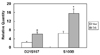
The relative DNA level of D21S167 in the blood was higher in Down syndrome subjects compared with the control group (6.16 versus 2.32). The relative quantity of S100B in the blood of Down syndrome subjects was also higher when compared with the control group (15.62 versus 6.47). The relative DNA levels of D21S167 and S100B were 2.6 and 2.4 times higher in the blood of Down syndrome patients than in the control group. The difference of DNA levels between these two groups was statistically significant (p-values: 0.0012 and 0.0016, Fig. 1).
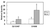
The relative DNA level of D21S167 in the amniotic fluid of Down syndrome fetuses was higher than that of the control group (6.37 versus 3.00). The relative DNA level of S100B in the amniotic fluid of Down syndrome fetuses was also higher when compared with the control group (19.14 versus 7.04). The relative DNA levels of D21S167 and S100B were 2.1 and 2.7 times higher in the amniotic fluid of Down syndrome fetuses than those in the control group. The difference of DNA level between the Down syndrome group and the normal control group was statistically significant (p-value 0.0379 and 0.0379, Fig. 2).
DISCUSSION
Currently, trisomy 21 can be detected using molecular genetic techniques such as polymerase chain reaction (PCR) or fluorescent in situ hybridization (FISH). One such method for detecting trisomy 21 is PCR using the specific primers for chromosome 21. Keuren et al.15 have reported that the genetic marker D21S167 was located on 21q.11.2-q22.2, and that the genetic marker D21S11 was located on 21q21 of chromosome 21. Trisomy 21 was defined as either a triallelic signal or a diallelic pattern, whereas disomy was indicated by the heterozygote pattern observed by fluorescent PCR using the D21S167 and D21S11 markers.10,11
This method was applied to other trisomies such as trisomy 18 and trisomy 13, using the D18S51 genetic marker for trisomy 18, and the D13S631 and D13S258 genetic markers for trisomy 13. Other trisomies have also been successfully detected.10
The S100B gene has been used for quantitative PCR to detect the DNA levels of chromosome 21.8 D21S11 and S100B were useful markers for the detection of trisomy 21 by quantitative PCR, and the prenatal diagnosis of trisomy 21 by PCR-associated STR analysis of S100B proved a useful, innovative, accurate and rapid method for diagnosis.9 In the analysis of S100B, the ratio of S100B to IGF-1 was 1.4:1.6 in Down syndrome samples, while the ratio of S100B in all the normal samples was close to 1:1 (Data not shown).
Quantitative polymerase chain reaction was introduced to measure the concentration of fetal DNA in maternal plasma and serum.16-21 Lee et al.12 have reported that the human liver-type phosphofructokinase gene located on chromosome 21 (PFKL-CH21) at position q22.3 in trisomy 21 is 1.5-fold higher by homologous gene quantitative PCR (HGQ-PCR) than that of PFKM-CH1 located on chromosome 1, and PFKM-CH10 located on chromosome 10. These results gave the first indication that this quantitative method could be used for the prenatal diagnosis of Down syndrome caused by trisomy 21.
Honda20 found that fetal gender was determined by conventional PCR by detecting a Y-chromosomal sequence (DYS14) in maternal blood with 95% sensitivity, whereas in real-time quantitative PCR, the total sensitivity after the fifth week was 100%. Bianchi22 reported that large amounts of cell-free fetal DNA were present in amniotic fluid when using real-time quantitative PCR of fetal DNA in the amniotic fluid. Bianchi found 0 GE/ml of the Y-DNA in the amniotic fluid for the female fetus, whereas the mean amount of male DNA in the male fetus was 3427 GE/ml. Lo et al.23 demonstrated that fetal DNA was present in maternal serum and plasma, and that the detection of fetal DNA sequences was made possible by recent developments in molecular biology. This would provide physicians and researchers a new screening test for fetal chromosomal disorders.
The recent introduction of real-time quantitative PCR, which is the most sensitive method for DNA and RNA detection, makes possible the rapid and accurate quantitation of specific DNA and mRNA. This system has a number of advantages: a high throughput and fast turnaround time, an accurate target quantitation over a wide concentration range within three to four hours, low cost, and it is possible to optically monitor the process continuously in the laboratory.
This study was designed to use D21S167 on 21q22.2 and S100B on 21q22.3 of chromosome 21 as targets of PCR. The blood of Down syndrome patients and the amniotic fluids of mothers conceiving Down syndrome fetuses were analyzed and compared with the normal control groups by real-time quantitative PCR. Based on the observation that D21S167 and S100B levels are high in the blood of Down syndrome patients, this study was designed to show that the levels of D21S167 and S100B in the amniotic fluid of Down syndrome patients are higher than in normal control groups. For our first attempts, 1 ng of DNA from blood and amniotic fluid was used as a template for PCR.
However, in the amniotic fluid, target genes were not amplified because the amount of DNA was too small. Therefore, we next used 100 ng of DNA from the amniotic fluid, which produced a good result.
The prenatal diagnosis of trisomy 21 by realtime quantitative PCR using STR (small tandem repeats) amplification of D21S167 and S100B is a useful, accurate and rapid diagnostic method. This method can also be employed in the diagnosis of trisomy 13 and 18. Furthermore, it may be useful for prenatal diagnosis using fetal DNA from maternal blood, and for preimplantation genetic diagnosis and prenatal counseling.
 XML Download