

 PDF
PDF ePub
ePub Citation
 Print
Print


An Arc of Buhler (AOB) is a persistent embryonal ventral anastomosis between the superior mesenteric artery (SMA) and the celiac axis (1, 2). The most common and important anastomoses between the SMA and celiac axis are the pancreaticoduodenal artery arcade (PDAA) and the dorsal pancreatic artery. An AOB is considered rare and independent of both the gastroduodenal and dorsal pancreatic artery (3). Furthermore, aneurysms of the AOB are even more uncommon, and are associated with stenosis or occlusion of the celiac axis, as seen in patients with PDAA aneurysms.
CASE REPORT
A 41-year-old male patient was referred by his primary physician for consultation regarding a visceral aneurysm detected from an abdominal CT. The patient frequently complained of mild dyspepsia and abdominal bloating. Moreover, the patient's medical history included medical treatment for acute pancreatitis about 10 years ago. While the patient was admitted to a local clinic for medical treatment, no radiologic examinations were performed at the time. Furthermore, the patient had no history of surgery, major trauma to the abdomen, or family history of aneurismal disease.
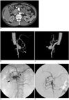
An axial CT angiography detected an aneurysm (Fig. 1A), which was later determined to originate from an anastomosis between the celiac axis and SMA following a 3-dimensional CT angiography (Figs. 1B, C).
Selective SMAs, using a 5-Fr Yashiro catheter (Terumo, Tokyo, Japan), showed a normal gastroduodenal artery, PDAA, and an anomalous communication, to form the AOB formation (Fig. 1D). In addition, an occlusion occurred at the celiac axis, which resulted in the retrograde filling of the splenic and common hepatic arteries from the SMAs, via the PDAA and gastroduodenal artery.
The AOB arose from the anterior surface of the proximal SMA and passed superiorly to join the proximal portion of celiac axis. This resulted in a 2-cm aneurysm with a wide neck at the origin of the AOB.
A 5-Fr Yashiro catheter was placed at the origin of the AOB, along with a 0.016-inch microguide wire (Conic 16 torque wire guide, Cook Inc., Bloomington, IN), which was passed into the AOB beyond the neck of the aneurysm. In addition, a microcatheter (Microferret 18, Cook Inc., Bloomington, IN) was passed over the microguide wire to the origin of the celiac axis. We decided to treat the aneurysm itself by a transcatheter embolization. The outflow segment was also treated by embolization through the same catheter, using Nester coils (Cook Inc., Bloomington, IN), and originating from the celiac axis to prevent reperfusion of the aneurysm sac. Next, the microcatheter was retracted from the outflow segment, transferred into the aneurysm itself, and packed with the same coils. Following the dense packing of the aneurysm with coils measuring: 14 cm × 10 mm (15), 14 cm × 6 mm (6), 14 cm × 4 mm (3), and 14 cm × 3 mm (1). The angiogram revealed a complete occlusion of the aneurysm (Fig. 1E) with preservation of the retrograde blood flow through the PDAA, as well as into the liver and spleen. The patient's hospital stay lasted just two days. Six weeks after the procedure, a follow-up abdomen CT angiography was performed, which revealed the exclusion of the aneurysm with a patent PDAA, as well as a normal perfusion into the liver and spleen with no detectable infarction. A subsequent patient follow-up after 10 months showed no symptoms of mesenteric ischemia.
DISCUSSION
The AOB was first described by Buhler and Tandler (1, 2) in 1904 as a vertically coursing artery, which directly connects the celiac axis and SMA. This anastomotic artery is independent of the gastroduodenal artery and the dorsal pancreatic artery. Moreover, it is diagnosed as the failure of regression at the ventral segmental anastomosis between the 10th and 13th segmental arteries during the embryonicstage.
The symptom manifestation in patients with aneurysms may occur at the celiac axis or at the SMA stenosis. A few patients with aneurysms are asymptomatic, with diagnosis occurring incidentally on CT, ultrasound imaging, celiac axis, or SMA angiography. Previous studies found that the incidence of AOB ranges from 1 to 4% in patients undergoing an arteriography for mesenteric arterial stenosis (symptomatic) (3, 4) and 3% in patients undergoing a mapping arteriography for a potential living-related liver transplant donation (asymptomatic) (5).
The AOB is anatomically associated with PDAA aneurysms, which are relatively uncommon, with slightly over 100 cases reported in the English-language literature. The incidence of PDAA aneurismal ruptures is reported in > 60% of cases cited in the present literature, with a mortality rate of 29% (6). No correlation was found between aneurysm size and the occurrence of ruptures, thus resulting in a strong recommendation for treatment of all unruptured aneurysms.
Aneurysms of the AOB are very rare, with only three cases reported to date. The first is by Kugai et al. (7), who described the treatment of a 3-cm saccular AOB in a patient with a celiac artery occlusion. Surgery was performed because a transcatheter embolization of the aneurysm was unsuccessful. The second AOB aneurysm case was described by Myers et al. (8), who reported the surgical resection of an AOB aneurysm with SMA to the splenic artery bypass, using the saphenous vein in a patient with von Recklinghausen's disease. Lastly, and most recently, Dubel et al. (9) described the successful transcatheter embolization of a 2.5-cm aneurysm of the AOB via a celiac stenosis and a patent PDAA.
Currently, the endovascular therapy of aneurysms is the preferred therapeutic method, due to its lower rate of morbidity and mortality. This approach involves the selective catheterization of the SMAs and then superselective catheterization of the AOBs. The aneurysm and outflow vessel are occluded with detachable and Nester coils, as was observed in the present case study.
The revascularization of a celiac stenosis, as the primary therapy for PDAA aneurysms has been reported (10), since the slow flow through the aneurysm was found to induce thrombosis within the aneurysm. Revascularization can be performed by an angioplasty and by stenting of the celiac axis. Alternatively, a bypass graft can be created between the celiac axis and the SMA. In the present case study, the celiac axis was occluded at the orifice, hence recanalization was not attempted.
In conclusion, the endovascular therapy of an asymptomatic aneurysm of the AOB with celiac axis occlusion and an intact PDAA, is a feasible and safe treatment method, based on our experience and previous case reports.
 XML Download