

 PDF
PDF ePub
ePub Citation
 Print
Print


Milk of calcium in the breast is well known as a benign entity identified in 4-6% of women that undergo diagnostic mammographies (1). Upon recognition, a short-term follow-up or biopsy can be avoided (1). However, carcinoma may incidentally arise adjacent to milk of calcium. To the best of our knowledge, the previously described carcinomas with milk of calcium were located near or adjacent to milk of calcium, not within them (2, 3).
We report a case of carcinoma presented as malignant microcalcifications mixed within milk of calcium in a breast.
CASE REPORT
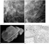
A 48-year-old woman underwent a screening mammography. The crainocaudal and mediolateral oblique views divulged a cluster of microcalcifications in the right upper inner quadrant. Subsequent magnification mammograms revealed the poorly defined smudges on a craniocaudal projection, and the typical crescent-shaped sedimented calcium on a mediolateral projection, also known as the "teacup sign", suggestive of milk of calcium (4). However, among the milk of calcium clusters, several fine pleomorphic microcalcifications, which are distinct in appearance from sedimented calcium, were observed (Figs. 1A, B).
And a mammography-guided wire localization a surgical excision was performed, followed by a specimen radiography, confirmed the excision of the calcifications (Fig. 1C). A histological assessment revealed a ductal carcinoma in situ, with a comedo type and a low nuclear grade among the cystically dilatated acini. The mammography initially revealed what was believed to be milk of calcium (Fig. 1D).
DISCUSSION
Milk of calcium has been found in a variety of cystic structures elsewhere in the body, particularly in the renal collecting systems and the gallbladder. The pathophysiology of milk of calcium formation is poorly understood; however, Lanyi (4) and Sickles and Abele (1) initially identified and explained the mammographic findings of milk of calcium in benign breast cysts. Because of its tendency to sediment out of cyst fluid, the diagnosis of milk of calcium diagnosis depended upon the demonstration of a poorly defined smudgelike density on a craniocaudal mammography as well as a fluid-calcium level on a true lateral view (1). Its recognition is important since this characteristic calcification has no known precedence as a malignant potential and a follow-up or biopsy has been typically deemed unnecessary (1).
However, as shown in our case, it may incidentally coexist with other types of calcifications, including malignant calcifications. In 1989, Linden and Sickles (2) described eight patients with carcinoma presented as clustered microcalcifications in a breast, which incidentally contained typical sedimented calcifications. Impriaco et al. (3) also described a similar case in 1999. The described cases revealed calcifications which were associated with carcinomas, were distinct in appearance from the sedimented calcium, but located near or adjacent to them however, not within them.
In the present case study, malignant microcalcifications were found to be located within the milk of calcium cluster. This finding brings attention to suspicious microcalcifications which are typically missed or are misdiagnosed.
The histology revealed that almost all calcifications were associated with malignant cells, but adjacent cystically dilated acini contained little calcific debris. From the mammographies, the calcific debris was identified as milk of calcium and hence, escaped the benign cyst lumen during the sectioning, as well as the transfer to glass slides, and staining. Kopans et al. (5) described 10 cases of typically benign calcifications associated with malignant cells, including milk of calcium layering in the dependent portion of cystically dilated acini lined with malignant cells. Even so, the classic crescent-shaped calcium precipitated in the cysts, and the spherical centrally lucent calcium deposits are almost always associated with the benign processes. The authors also believe that it was the malignant calcifications seen at histology that were the fine pleomorphic calcifications seen mammographically, and not milk of calcium.
As a result of this case study, radiologists should be aware of the possible association between milk of calcium and other types of calcifications, including malignant calcifications. A more careful analysis of the characteristics of all calcific particles and correlation with craniocaudal mammograms with true lateral views is essential. Unless all calcific particles are characteristic of a benign entity, the consideration of a biopsy would be a prudent approach.
 XML Download