

 PDF
PDF ePub
ePub Citation
 Print
Print


Anisakiasis is a parasitic infection caused by the consumption of raw or insufficiently pickled, salted, smoked, or cooked wild marine fish infected with larval nematodes belonging to the family Anisakidae (1). Anisakiasis is most commonly associated with the stomach wall and small intestines. The colon has only rarely been found to be affected (2). The clinical diagnosis of colonic anisakiasis is very difficult due to the nonspecificity of the symptoms and the rarity of the condition. The majority of cases are initially misdiagnosed as appendicitis, cancer, and inflammatory bowel diseases such as Crohn's disease or intestinal tuberculosis (3-5). Furthermore, the differential diagnosis becomes even more difficult when the colonic anisakiasis is combined with a cancer lesion (6). Only one case of colonic anisakiasis associated with a carcinoma in the colon has ever been reported (6).
The CT colonography (CTC) has emerged as a valid diagnostic test for colorectal cancer (7). As CTCs are less invasive and compare favorably with a colonoscopy in the detection of colorectal polyps 6 mm in diameter or larger, it has rapidly gained popularity (7). Furthermore, this has led radiologists to identify adenomatous or hyperplastic polyps, as well as other colonic lesions, such as diverticulosis, submucosal tumors, infection, and inflammatory disease (8-10).
We present a case of synchronous colon cancer and colon anisakiasis on a CTC.
CASE REPORT
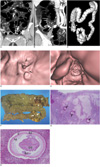
A 50-year-old woman was admitted to our hospital complaining of severe left lower quadrant pain which began 10 days prior to admission. The patient experienced one episode of hematochezia and a change in stool caliber, both of which first began two months before. The patient also had a history of hypertension and an appendectomy. A physical examination identified tenderness in the right lower quadrant of the abdomen. The laboratory findings, upon initial admission, showed a slight decrease of hemoglobin (11.5 g/dL) and hematocrit (33.1%) level, as well as a marked elevation of the eosinophil count (17.1%). The serum level of the carcinoembryonic antigen (CEA) was within the normal range. An abdominal plain radiography revealed a distension of the bowel loops, which was a sign of ileus. A coronal multiplanar reconstruction image of a CTC demonstrated a concentric and enhancing wall thickening, along with luminal narrowing in the sigmoid colon (Fig. 1A), and a low attenuating wall thickening with pericolic infiltration in the ascending colon (Fig. 1B). A volume-rendered surface-shaded image of a CTC showed an apple-core shaped, concentric narrowing in the sigmoid colon (Fig. 1C). In addition, a segmental fold thickening with a thumbprint-like appearance (Fig. 1C) was seen in the ascending colon. A virtual endoscopic image of the sigmoid colon revealed a concentric mass (Fig. 1D), which led to a narrowing of the colon. An irregular fold thickening and mild luminal narrowing (Fig. 1E) was also observed via a virtual endoscopic image of the ascending colon. A colonoscopy was performed; however, due to the severe obstruction of the sigmoid colon by the concentric mass, the scope could not traverse the mass and hence, could not attain the ascending colon.
The diagnosis of synchronous double primary colon cancers was made preoperatively. The patient underwent total colectomy, and a gross pathologic examination of the resected specimen (Fig. 1F), which showed an ulceroinfiltrative mass in the sigmoid colon and another ulcerative mass in the ascending colon. The adjacent folds around the ascending colonic mass were edematous. Histologically, the sigmoid mass was diagnosed as an adenocarcinoma infiltrating the subserosal layer. The ascending colonic wall was heavily infiltrated by eosinophils, in addition to the presence of several parasite worms in the submucosa (Fig. 1G). Moreover, diagnostic morphologic characteristics of sections in the intestinal region of the nematode were made (Fig. 1H). In addition, a thin external cuticle with no lateral alae and a muscle layer with prominent Y-shaped lateral epidermal cords were observed. The digestive tract of the parasite consisted of a single layer of columnar epithelial cells with no apparent reproductive system. A pathologist confirmed this lesion as anisakiasis. According to the patient's account, she frequently consumed raw marine fish at Japanese restaurants. The final diagnosis of human colonic anisakiasis associated with a carcinoma was based on the morphology of the parasite and the frequent history of raw marine fish consumption. The preoperative findings were not adequate to differentiate between advanced colonic carcinoma and anisakiasis in this case.
DISCUSSION
Anisakiasis was first described over 40 years ago in a patient with severe abdominal pain resulting from the ingestion of raw herring (11). Since then, thousands of cases have been reported worldwide, but the colon has only rarely been the site of involvement (11). Mineta et al. (11) performed a meta-analysis of as much as 30,000 reported cases of anisakiasis in Japan, and found only 75 cases of colonic anisakiasis by 2001. Among the 75 reported cases of colonic infection, more than half of the cases were on the right side (11). The most reasonable explanation for the rarity of this colonic infection is that the colon is too far for the orally ingested larvae to attain the colon (2, 11). The more frequent involvement of the right side of the colon can be explained by the gradual peristalsis and slow transit in the colon, which allows the larvae to infect while still in the right side of the colon (2).
In the case of colonic anisakiasis, symptoms may include diffuse abdominal tenderness or colicky abdominal pain, nausea, and vomiting (2, 3, 11-13). Because of these nonspecific symptoms, colonic anisakiasis is diagnosed unexpectedly after a histopathologic examination of surgical specimens resected for other reasons, particularly appendicitis or acute abdominal syndrome (11). In addition, colonic anisakiasis may simulate a tumor of the colon, since this infection provokes edema, an acute phlegmonous reaction, or granuloma formations around the larvae in the submucosa of the intestinal wall, which results in a mass effect (2, 14). Hence, the preoperative assessment also misdiagnosed this case as synchronous double primary colon cancers.
The radiographic features of intestinal or colonic anisakiasis are characterized as edematous changes of the enteric walls, including thickened folds, a "thumb-print" appearance, a saw-toothed aspect, an irregular narrowing of the lumina, and the disappearance of kerckring folds (1, 2). The imaging findings of anisakiasis on a CTC have never previously been described. The CTC has emerged as a valid diagnostic colorectal cancer test, and its role in the diagnosis of colorectal disease is being extended (7). As a CTC is a less invasive procedure and compares favorably with a colonoscopy for the detection of colorectal polyps with diameters of 6 mm or larger, it has rapidly gained widespread acceptance (7). In addition, a CTC is considered as an effective method for evaluating the entire colon region before surgery in patients with occlusive colorectal carcinomas (15). Approximately 1.5%-9% of patients with colorectal carcinomas have a synchronous cancer, whereas 27%-55% have multiple coexistent adenomatous polyps (15). Because a failure to identify synchronous carcinomas before surgery in patients with colorectal cancer leads to serious negative prognostic and therapeutic consequences, the preoperative evaluation of the entire colon in patients with colorectal cancer is strongly recommended. Although, the presence of occlusive carcinomas can preclude adequate evaluations of the proximal colon with an optical colonoscopy. Unlike a colonoscopy, a CTC provides a complete profile of the colon, and thus, improves the accuracy in the depiction of synchronous colorectal neoplasms, even in occlusive cancers (15, 16). The frequent use of the CTC has given the ability to radiologists to identify adenomatous and hyperplastic polyps, as well as other colonic lesions, such as diverticulosis, submucosal tumors, infection, and inflammatory disease (8-10). In these reports, signs suggesting the presence of inflammation on a CTC included, wall thickening involving a rather long segment of the colon, fold thickening, luminal distortion, and the flattening or disappearance of the haustra (8-10). Despite the initial clinical diagnosis being synchronous double primary colon cancers, from a retrospective review, we revised our assessment to incorporate the signs of inflammation as well as a colon cancer on a CTC. Our case demonstrated a segmental fold thickening with a thumbprint-like appearance in the ascending colon, which suggested inflammation rather than cancer, in contrast to the typical apple-core shaped, mucosal destructing, and concentric narrowing in the sigmoid colon cancer. Despite the correction of the diagnosis, the radiologic appearances are still nonspecific and too difficult to differentiate anisakiasis from other inflammatory or tumorous conditions solely through radiologic findings. Moreover, although peripheral eosinophilia may be considered as a diagnostic indicator for a parasitic infection, it sometimes lacks specificity (3). Eosinophilia and gastrointestinal symptoms may also be consistent with Crohn's disease, Hodgkin's lymphoma, and eosinophilic gastroenteritis (2). Instead, the most important clinical clue for the correct diagnosis of colonic anisakiasis should be a history of raw marine fish ingestion (3). In a retrospective interview, our patient reported her frequent history of consuming raw marine fish at Japanese restaurants.
To the best of our knowledge, these images represent the first reported visualization of synchronous colon cancer and colon anisakiasis on a CTC. Despite the more frequent occurrence of synchronous double colon cancers, the combination of colon cancer and a parasitic infection is on the rise due to the worldwide popularity of the consumption raw fish, and it is necessary for primary physicians to consider the possibility of associated parasitic infections when presented with heavy peripheral eosinophilia, a thumbprint-like fold thickening with little mucosal destruction on a CTC, and a history of consuming raw fish.
 XML Download