

 PDF
PDF ePub
ePub Citation
 Print
Print


Carney complex is a familial multiple neoplasia disorder that's characterized by cardiac and cutaneous myxomas, spotty pigmentation and endocrine overactivity (1-6). Female patients with Carney complex often have benign myxoid fibroadenomas and ductal adenomas in the breast (2, 3, 7). We experienced the case of a 37-year-old woman with increased myxoid fibroadenomas, and she showed the typical clinical findings of Carney complex. The clinical work-up for Carney complex identified multiple intracranial aneurysms.
CASE REPORT
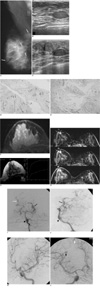
A 37-year-old woman presented with complaints of mild pain in both breasts. The mammogram showed multiple nodules in the right breast (Fig. 1A). On ultrasound (US), we identified 16 hypoechoic or isoechoic well-defined nodules in the right breast and seven in the left breast (Fig. 1B). US-guided biopsy was performed in both breasts and this proved the presence of myxoid fibroadenomas (Fig. 1D).
After about three months, the patient revisited the hospital with severe pain in both breasts. Follow-up US of the breast revealed increased hypoechoic or isoechoic well-defined nodules in both breasts (33 in the right and 14 in the left) (Fig. 1C). Magnetic resonance imaging (MRI) of the breast showed multiple well-defined ovoid nodules. These lesions had bright signal intensities on the T2-weighted images and they showed persistent enhancement on the T1-weighted dynamic MR images, which are both characteristics of myxoid fibroadenomas (Figs. 1F-H). Two 5 mm sized extruding masses on the right forearm and the right nostril, respectively, were found on physical examination. Her face had spotty pigmentation and lentiginosis. Carefully taking the past medical history revealed that she had cardiac myxomas excised at the age of 10 and 20. The two masses of the right forearm and the right nostril were excised and they were shown to be myxomas (Fig. 1E). The cardiac myxomas, mucocutaneous myxomas, spotty pigmentation and myxoid fibroadenomas in the breast led to the diagnosis of Carney complex. Yet she had no family history of this condition.
Brain MRI, US of the thyroid, abdomino-pelvic CT and laboratory studies were performed to evaluate for endocrinopathy such as pituitrary adenomas, thyroid adenomas and carcinomas, ovarian cysts and adrenocortical disease. The results showed no evidence of endocrinopathy. The brain MRI incidentally revealed abnormally dilated vascular structures on the right parietooccipital area and on the left perimesencephalic cistern. The scan protocol of brain MRI included the axial and sagittal T1-weighted (TR/TE 630/14) images, the axial T2-weighted (TR/TE, 3588/99) images and the contrast-enhanced axial T1-weighted images. Conventional four-vessel cerebral angiography was performed. It depicted multiple fusiform dilatations in the basilar artery, the proximal posterior inferior cerebellar artery, the left posterior cerebral artery (P2 segment), the right posterior cerebral artery (P4 segment), the temporal branch of the left middle cerebral artery and the distal branches of the right middle cerebral artery and the right anterior cerebral artery (Figs. 1I-L).
The patient will undergo follow-up cardiologic, neurologic and radiologic examinations at regular intervals.
DISCUSSION
Carney and colleagues first reported on Carney complex in 1985 as the complex of myxomas, spotty pigmentation and endocrine overactivity (1). This is transmitted in an autosomal dominant fashion and the disease has a familial pattern in approximately half of the patients; this disease appears sporadically in the remaining patients (3). The diagnosis is made when there is at least one characteristic clinical finding associated with the disorder and there is a family history of Carney complex or at least two characteristic clinical findings in the absence of a family history (4). Carney complex should not be confused with Carney syndrome/triad, which is a distinct neoplasia syndrome, and the characteristics of Carney syndrome/triad are gastric leiomyosarcoma, pulmonary chondroma and extra-adrenal paraganglioma (4).
Cardiac and cutaneous myxomas, pigmentation abnormalities and endocrine tumors are the main manifestations of this disease (5). Cardiac myxoma is an important manifestation of Carney complex. Unrecognized cardiac myxoma may be the cause of the sudden death due to embolization (4, 5). Pigmentation abnormalities are the most common feature (4). Lentiginosis is observed in most patients and this is so characteristic that it can be used to make the definite diagnosis (5). The common endocrine tumors of Carney complex are growth hormone secreting pituitary adenomas, thyroid adenomas and carcinomas, testicular tumors, ovarian cysts and Cushing's syndrome, and the latter is due to primary pigmented nodular adrenocortical disease (4, 5).
There are numerous manifestations of Carney complex and these vary between patients. In this patient, myxoid fibroadenomas in the breast were one of the main manifestations and in particular, the number of the lesions increased at an interval of three months. About a fifth of the women with Carney complex show abnormal, but generally benign breast pathology (2, 7). Myxoid fibroadenomas are the typical findings and they can be multicentric and bilateral, as in this case (3, 7). Ductal adenoma of the breast is also a recurrent finding in women with Carney complex (2, 3). These two diseases may coexist in the breast of patients with Carney complex (3). On the T2-weighted MR images, high-signal-intensity lesions are characteristic of myxoid fibroadenoma (3). If additional lesions with similar features exist and no other signs of malignancy are present, then the lesions should not prompt the physician to perform multiple biopsies and the lesions can be followed up clinically (3).
This patient has multiple fusiform intracranial aneurysms. There are several possible explanations for the association of Carney complex and multiple intracranial aneurysms. One possible explanation is that the cerebral aneurysms are related to Carney complex as a coincidental finding. However, numerous intracranial aneurysms are extremely rare in the normal population, making this explanation unlikely.
Another explanation is that multiple intracranial aneurysms develop as a delayed complication of cardiac myxoma. The association of cardiac myxoma and multiple intracranial aneurysms has rarely been documented. Though it is uncommon, myxomatous aneurysms may develop years after successful excision of the cardiac myxoma. The pathogenesis of these aneurysms seems to be that tumor material from the cardiac myxoma embolizes into the vasa vasorum of the peripheral arteries, and this subsequently proliferates in the vessel wall and weakens the subintimal tissue such as the internal elastic lamina (8, 9). The disrupted vascular wall then dilates in an aneurysmal manner. The typical angiographic findings include peripherally located, fusiform aneurysms (8, 9).
Third, intracranial aneurysm has been associated with various heritable connective tissue disorders. The most important diseases are Ehlers-Danlos syndrome type IV, Marfan's syndrome, neurofibromatosis type I, autosomal dominant polycystic kidney disease and α-1-antitrypsin deficiency fibromuscular dysplasia (10). Like these diseases, multiple intracranial aneurysms as a part of Carney complex might also be included as a potential explanation.
In conclusion, the manifestations of Carney complex can be numerous and they can vary between patients. Multiple and increased myxoid fibroadenomas in the breast can be a particular manifestation of Carney complex. In this case, the multiple intracranial aneurysms may also represent an additional finding of Carney complex.
 XML Download