

 PDF
PDF ePub
ePub Citation
 Print
Print


There have been about 20 case reports describing an aberrant right subclavian artery (ARSCA) and an anomalous origin of the right vertebral artery from the right common carotid artery (right VA-CC) (1-10). We recently experienced two cases of ARSCA with a right VA-CC, and these were confirmed by the enhanced CT images. In this study, we discuss the embryologic mechanism of this variation. Based on our cases, we evaluate the incidence of right VA-CC with underlying ARSCA and the clinical implication of this variation, and we especially focus on the arterial course of the vertebral artery.
CASE REPORTS
Case 1
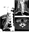
A 30-year-old male was admitted to our hospital due to a midline anterior neck mass. On the enhanced CT images, the mass was located at the infrahyoid midline neck and it showed the imaging features of a thyroglossal duct cyst. The enhanced oblique coronal multiplanar reconstruction CT images revealed a variation of the aortic arch as an aberrant right subclavian artery that was distal to the left subclavian artery (Fig. 1A). The right vertebral artery had its origin from the right common carotid artery at the inferior border of the right thyroid gland, and it had an aberrant entrance to the C5 transverse foramen (Fig. 1B). The prevertebral segment of the right vertebral artery was located in the retro-thyroid area and very close to the thyroid gland (Fig. 1C).
Case 2
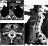
A 67-year-old female was admitted to our hospital for an operation for thyroid cancer. The enhanced CT images revealed an aberrant right subclavian artery distal to the left subclavian artery (Fig. 2A). In addition, the left vertebral artery originated from the aortic arch between the left common carotid artery and the left subclavian artery. The right vertebral artery had an origin from the right common carotid artery and also an aberrant entrance to the C5 transverse foramen (Fig. 2B). The left vertebral artery also had an aberrant entrance to the C5 transverse foramen. The prevertebral segment of the right vertebral artery was located in the retro-thyroid area and close to the thyroid gland (Fig. 2C).
DISCUSSION
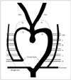
The embryologic mechanism of ARSCA with a right VA-CC has been explained in the several studies (2, 4, 8). The normal vertebral artery (VA) builds up due to the process of longitudinal anastomosis and obliteration of the horizontal parts of the cervical intersegment artery. Normally, the first to the sixth cervical intersegment arteries (CIAs) develop into the VA and the seventh CIA makes the subclavian artery (SCA). If longitudinal anastomosis of the right CIA stops between the 6th and 7th CIA, and the right side of the dorsal aorta is obliterated proximal to the 7th CIA, then the right side subclavian artery (SCA) originates from the left side aorta distal to the left SCA and the right VA originates from the right common carotid artery (Fig. 3).
Aberrant right subclavian artery has been reported with the incidence of less than 1% (8). The known incidence of right VA-CC is about 0.18% (1). A combination of these two variations is rare, but the true incidence of right VA-CC with underlying ARSCA is not known. Fifteen cases of ARSCA were confirmed by enhanced CT or CT angiography in our hospital during the recent three years. Among these cases, only the two cases we present herein showed the right VA-CC variation. Also, only these two cases had an aberrant level of the entrance of the VA into the transverse foramen of the cervical spine. Based on our results, the right VA-CC variation is not likely to frequently occur with an underlying ARSCA.
There have been many reports about the variation of the ARSCA with a right VA-CC, but only three reports remarked about the aberrant entrance of the VA into the transverse foramen of the cervical spine (3, 7, 10). The vertebral artery usually enters into the transverse foramen of the 6th cervical spine (the C6 entrance). Those three reports revealed different entrance levels as C2, C3 and C4. In our two cases, the right VAs had a C5 entrance. There have been no reports describing the anatomical course of this type of VA-CC. In our cases, this VA-CC had a close spatial relation with the thyroid gland. It was located in the retro-thyroid area and very close to the thyroid gland. During its course up to the transverse foramen, it traveled above the longus colli muscle. This anatomical characteristic of the VA-CC bears watching during anterior cervical spine surgery, thyroid surgery or other interventions. If this VA-CC were overlocked, it may be pulled with the longus colli muscle, or it may become lacerated during the anterior crevical spine surgery. During thyroidectomy, the inferior thyroid artery is usually ligated. As mentioned by one autopsy result (7), the inferior thyroid artery may be near to the right VA-CC, so meticulous care may be needed to avoid an inadvertent injury to the VA-CC during thyroidectomy. During thyroid aspiration, the needle occasionally penetrates the posterior surface of the thyroid gland and it reaches the vertebral body. If the VA-CC is near to the thyroid gland, then there is a possibility to puncture the VA during thyroid aspiration. Therefore, knowledge of this aberrant course of the VA may be helpful to avoid injury of the VA when performing these procedures.
 XML Download