

 PDF
PDF ePub
ePub Citation
 Print
Print


Cholesterol granuloma (CG) is a histopathological term used to describe numerous clefts that form after cholesterol crystals have dissolved during processing, foreign body giant cells, foam cells and macrophages filled with hemosiderin that are embedded in fibrous granulation tissue and often exhibit fibrin deposition and focal bleeding (1-5). Although CGs can develop in the middle ear, mastoid bone, paranasal sinus, breast (6) and sella turcica, their occurrence in the paranasal sinuses is very rare (1-5, 7). However, when CG does occur in the paranasal sinus, it is most commonly found in the maxillary sinus, although one case of CG in the sphenoid sinus has also been reported (8). In addition, to the best of our knowledge, there has been only one reported case involving the association of CG and aspergillosis in the paranasal sinus (1).
The most frequent imaging findings of CG in the paranasal sinus are the presence of opaque antrum or cystic lesions with soft tissue density and suspected bony destructions (9), and most cases of CG have been treated using radical operative techniques (7).
This paper describes a case of a sphenoid sinus CG that was associated with an aspergilloma. In addition, the imaging characteristics of the CG observed upon computed tomography (CT) and magnetic resonance imaging (MRI) are also described herein.
CASE REPORT
A 78-year-old male patient presented with complaints of right hemifacial pain that had persisted for one year, as well as a headache and toothache that had been present for one week. The patient's general history revealed hypertension and diabetes mellitus, however, there were no abnormal findings detected during the physical examination.
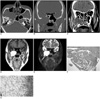
CT revealed the presence of a 4.8 × 3.7-cm soft tissue density mass lesion in the right sphenoid sinus that extended to the right infratemporal and pterygopalatine fossae. In addition, CT revealed that the right infratemporal and pterygopalatine fossae, as well as the right pterygoid process and the posterior wall of the right maxillary sinus showed indentation and erosive changes as a result of the mass lesion (Figs. 1A, B). Further, the CT scan revealed sclerosis and thickening of the bony wall of the right sphenoid sinus (Fig. 1B), as well as a tiny calcified lesion at the inferior portion of the mass lesion (Fig. 1C).
Upon MRI, the mass lesion was shown to have a high signal intensity on both the T1 weighted (T1WI) and T2 weighted images (T2WI) (Figs. 1D, E), and another 10 × 7-mm-sized high T1 and low T2 nodule was seen at the inferior portion of the mass lesion (Figs. 1D, E).
Based on these findings, it was assumed that this mass was a right sphenoid sinus tumor, such as a CG or an inspissated proteinaceous mucocele, that extended into the right pterygopalatine and infratemporal fossae. Therefore, the tumor was excised using a transmaxillary approach. The surgical specimen was then submitted for pathological examination, which revealed that it consisted of loose edematous and friable grey-white soft tissue.
The tumor was also examined microscopically and found to contain spindle-shaped cholesterol clefts surrounded by foreign body giant cells and granulation tissue formation. In addition, abundant erythrocytes and foamy histiocytes were also found to be present in the mass (Fig. 1F). Moreover, a separate fragment comprised of mycelium with septate hyphae that showed dichotomous branching was also observed (Fig. 1G).
Based on the results of the histopathological analyses, diagnoses of chronic sinusitis, CG and aspergilloma were made.
DISCUSSION
Cholesterol granuloma is usually observed in the middle ear or in cases involving temporal bone diseases, and is rarely seen in the petrous apex, orbit or paranasal sinuses (1-5, 7). The etiologic mechanism by which CG formation occurs is not well understood, however several studies have suggested that they occur as a result of hemorrhage, impaired drainage, and obstruction of ventilation (1, 3-5). One possible mechanism by which CG may occur is as a result of chronic sinusitis or previous surgery that causes an obstruction in the sinus ostium, which in turn results in the resorption of gas in the obstructed cavity. This resoprtion of gas then creates a relative vacuum that leads to the dilated mucosal blood vessel rupturing. Erythrocyte cell membranes that are destroyed during bleeding then act as a source of cholesterol, which is formed into cholesterol crystals by anaerobic erythrocytes. These crystals then incite foreign body giant cell infiltration, and repeated hemorrhaging ultimately leads to the formation of granulation tissue (1, 3-5).
Imaging findings of a CG in the paranasal sinus described in a previous study were seen as opaque antrum or cystic lesions with soft tissue density (9). In general, the morphology of a CG shows an expansile nature with scalloping of the surrounding bone; however CG must be differentiated from mucoceles and cystic schwannoma. Although cystic schwannoma can be excluded based on characteristic imaging features, it may be difficult to differentiate CGs from mucoceles based on CT analysis (5) because both appear as areas of near-fluid attenuation of expansile lesions with thinning of the adjacent bone and erosion. However, upon MRI, CGs show a high signal intensity on the T1WI due to the paramagnetic effect of methemoglobin, and they also show a high signal intensity on the T2WI as a result of the granulation of the paranasal sinus. Therefore, MRI can be used to differentiate CGs from mucoceles. In this case, the MR imaging characteristics of the CG is well described, however, inspissated proteinaceous mucoceles or internal hemorrhages can also show high signal intensity on the T1WI, therefore, a histopathological diagnosis may occasionally be necessary (5).
Histopathological observation of the CG revealed a cholesterol crystal cleft surrounded by granulation, as well as infiltration by multinucleated giant cells. CGs are composed of cholesterol, which is a component of cell membrane-producing steroid hormones and bile acids. When cells, including erythrocytes, are destroyed, the cholesterol and cholesterol esters that compose the CG are released, and these compounds then precipitate in crystalline form. Precipitation of the cholesterol then leads to an inflammatory reaction that induces the migration of macrophages and inflammatory cells, forming granulation tissue (1, 4-6).
To our knowledge, the association of CG with aspergillosis is extremely rare (1, 10). Aspergilloma is a form of fungal sinusitis that is characterized by the presence of tightly packed fungal hyphae with calcium phosphate and sulfate deposits within the necrotic mycetoma that are visible upon microscopic analysis. Aspergilloma also usually show hypointensity upon T1WI as a result of the absence of free water in the thick, solid mass; however, the signal intensity can be very heterogeneous. In addition, aspergilloma show low signal intensity on the T2WI, which may be due to macromolecular protein binding (11, 12). Cases of paranasal aspergilloma, such as the one described in this paper, may be associated with chronic sinusitis (1). This is because chronic sinusitis can result in impaired ventilation and drainage as a result of ostia obstruction and mucosal swelling, which favors fungal infection (1).
Although CG and aspergilloma appear to have similar etiologic mechanisms, such as impaired drainage and obstruction of the paranasal sinuses, there is no causative relationship between CG and aspergilloma. Further, the breakdown products of the fungi cannot be the source of the cholesterol granuloma because the major sterol in the plasma membrane of fungi is not cholesterol, but ergosterol (1), and there have been no reports of granulation formation as a result of ergosterol deposition (1).
Overall, in this paper, we describe a case of CG that was associated with aspergilloma that occurred in the sphenoid sinus, but was extended to the infratemporal and pterygopalatine fossae. The association observed in this case appears to be the result of the similar pathogenesis of these two diseases.
 XML Download