

 PDF
PDF ePub
ePub Citation
 Print
Print


Leiomyosarcoma is a malignant tumor arising from the smooth muscle cells. Although it may involve any organ in the body, involvement of the vessel wall is rare. Vascular leiomyosarcomas represent less than 2% of all leiomyosarcomas (1). The inferior vena cava is the most commonly affected vessel, accounting for 60% of all cases involving the vessel wall (2). The large central veins and the long saphenous veins are the next most commonly affected areas (1, 2).
A few sporadic cases of leiomyosarcoma of the ovarian vein have been described in previous reports (2-5). Among the previously reported cases, three cases showed abnormal excretory urographic findings (2, 4, 5), and two cases were confirmed by computerized tomography (CT) (4, 5). However, conventional CT was used only to confirm the presence of the retroperitoneal mass below the kidney. There was no mention that CT was able to preoperatively identify that the origin of the tumor was vascular (4, 5). In the present case, multi-detector CT and ultrasonography identified the retroperitoneal mass with intravascular growth patterns along the anatomical site of the right ovarian vein, and confirmed the diagnosis preoperatively. However, the use of ultrasonography had some limitations. We also discuss the imaging findings of ovarian vein leiomyosarcomas in comparison with other venous or retroperitoneal leiomyosarcomas.
CASE REPORT
A 39-year-old woman visited the hospital and presented with a hard, firm and palpable mass in the right upper abdomen. The medical and family histories were not remarkable. Serum levels of the tumor markers, including CA 19-9, carcinoembryonic antigens and CA 125, as well as laboratory findings, were all normal.
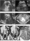
Excretory urography revealed lateral displacement of the lower pole of the right kidney and ureteropelvic junctional obstruction caused by the space-occupying lesion. An abdominal ultrasonogram (Acuson Sequoia 512; Siemens Medical System, Erlangen, Germany) showed the presence of a solid medium-echoic retroperitoneal mass with partial invasion of the inferior vena cava (Figs. 1A-C). An examination with 16-channel multi-detector CT (Mx8000 IDT; Philips Medical System, Eindhoven, the Netherlands) revealed the presence of a lobulated and well-defined right retroperitoneal mass measuring 5.1×5.5×6.9 cm with extensive cystic degenerations and highly enhanced solid portions on contrast-enhanced scans (Fig. 1D). Coronal images definitely confirmed the vertically elongated arm-like structures contiguous with the mass, extending upwards to the right renal vein with partial protrusion into the inferior vena cava (Fig. 1E). In the downward direction, the mass was connected to the normal right ovarian vein in the pelvis (Fig. 1F). The mass seemed to be separated from the adjacent right kidney and small bowels. Both renal and iliac angiograms showed the tumor vessels originating from the inferior segmental branch of the right renal artery.
During surgery, the mass was found to be located in the retroperitoneal space inferomedial side of the right kidney. The upper portion of the right ovarian vein was distended with tumor thrombi (Fig. 1G). There was no invasion of the adjacent organs and no enlarged lymph nodes.
Crosscut sections of the tumor showed mainly solid, and partly hemorrhagic and necrotic appearances with a yellow-tinged gray color. Microscopically, the tumor showed a typical pattern of intersecting fascicles of spindle cells with hyperchromatic nuclei and eosinophilic cytoplasms, representing the smooth muscle cells in the media. The tumor cells frequently showed highly pleomorphic nuclei, indicative of dedifferentiation. Immunohistochemically, the tumor cells were strongly positive for both smooth muscle actin and desmin.
The cells showed positive expression of CD34. Expression of CD117 (KIT), usually expressed in gastrointestinal stromal tumors and normally negative in leiomyosarcoma, was negative.
Adjuvant radiotherapy was performed over a six-week period. Follow-up abdominopelvic CTs after two and three months showed the presence of a 1.0 cm low attenuation nodule in the liver, that had newly developed and enlarged in size over the short-term intervals. We assumed the presence of a liver metastasis, but the patient was lost to further planned follow-up.
DISCUSSION
Leiomyosarcomas of the vein arise from the smooth muscle cells of the tunica media of the vessel wall. They grow slowly and bilaterally along the wall of the vessel. Retroperitoneal leiomyosarcomas exhibit themselves as completely extravascular (62%), completely intravascular (5%) or with both extra- and intraluminal growth patterns (33%) (6). As the leiomyosarcomas of the inferior vena cava grow, they eventually invade adjacent tissues and metastasize distantly; however, they tend to expand along the tissue plans of least resistance. There are no reported cases of tissue invasion of the adrenals, kidneys or bowels for leiomyosarcomas of the inferior vena cava (7). Like other retroperitoneal tumors, these tumors seldom present with symptoms until they progress into a huge mass. When the tumors are discovered at this later stage, there is a poor outlook for long-term survival.
As the cases are rare, previously reported radiological findings of a ovarian vein leiomyosarcoma are few in number. The lesions have been merely described as a heterogeneous retroperitoneal mass with ipsilateral hydronephrosis by conventional CT and excretory urography (2, 4, 5).
The findings of excretory urography, representing the effect of mass, are not specific for the diagnosis of the leiomyosarcomas of vascular origin.
The general findings of gray-scale ultrasonography of an inferior vena cava leiomyosarcoma are solid but often show cystic necrosis with irregular walls (6, 8). In our case, the sonographic findings were compatible with those of the CT scans, i.e., the pedicular structure was a tumor-invading ovarian vein. However, sonography had some limitations in that it failed to demonstrate the whole features of the tumor and the relationship between the tumor and the normal ovarian vein in the pelvis. These limitations may be due to the overlying bowel, the extent of the mass, or limitations in the depth resolution of the ultrasound modality (6).
The use of CT is superior to the use of sonography to reveal the highly enhancing and necrotic characteristics of the tumor, which is specific for the diagnosis of leiomyosarcoma (7). When CT shows a solid or necrotic extraluminal retroperitoneal mass not originating from a retroperitoneal organ, and possessing a contiguous intravascular enhancing appearance, it is accepted as a pathognomonic finding of vascular leiomyosarcoma (6). Low-attenuation areas representing necrosis, or rarely, a cystic mass due to diffuse necrosis, may be seen (7, 9). In our case, the eccentric cystic area is visible on sonography and CT; however, the extensive central necrosis is only visible on CT.
With great advances in digital imaging and high spatial resolution of multi-detector CT over the last few decades, scrolling through multi-stacked images and conducting multiplanar reconstruction is possible. These methods are useful in demonstrating the relationship between a mass and vessels, as well as detecting the tumor or venous thrombi. Thus, it was not difficult to presume that the mass originated in the right ovarian vein based on anatomical conceptions, as the tumor possessed vertically elongated bi-armed structures, the typical site of the right ovarian vein.
The angiographic findings of vascular leiomyosarcomas are generally accepted as nonspecific for the diagnosis. Angiography may have a role for demonstrating the primary vessels supplying the tumors and determining the degree of vascularity (6, 9).
Metastases will occur eventually in most survival cases of other venous or retroperitoneal leiomyosarcomas (1, 2, 6). Hematological metastasis occurs more commonly than lymphatic metastasis. The frequent sites for metastases are the liver, lung, brain, and peritoneum (1, 2, 6, 8). Local recurrence occurs occasionally in 40% to 77% of cases, even though a complete excision was made (6). Our case also showed the CT findings of a liver metastasis, although a confirmation was not obtained.
In conclusion, the general imaging findings of our case were coincidental with those of other venous or retroperitoneal leiomyosarcomas. Sonography seemed to be helpful for the diagnosis of ovarian vein leiomyosarcoma, but the use of multi-detector CT was the tool of choice to confirm the diagnosis. Scrolling through multi-stacked images and multiplanar reconstruction of the CT images have great benefits for the preoperative predictions of the location, extent, and growth patterns of the tumor, as well as the relationship of the tumor and vessels. We expect that the primary diagnostic tool for vascular leiomyosarcomas will be the use of multi-detector CT and it may expel the other unnecessary, cost consuming or invasive diagnostic procedures.
 XML Download