

 PDF
PDF ePub
ePub Citation
 Print
Print


Asolitary fibrous tumor (SFT) is a rare neoplasm, which is traditionally presented as a pleural-based mass, but can also occur in unusual locations based on its mesenchymal origin (1-4). An SFT arising from the larynx is very rare; only six cases of this tumor have been reported (5-9). Moreover, only limited descriptions of the imaging features of this rare tumor involving the larynx appear in the literature.
Herein, we report a case of an SFT of the larynx, with computed tomography (CT), magnetic resonance (MR) imaging, and angiography findings, along with its pathologic correlation.
CASE REPORT
A 34-year-old man was referred to our hospital because of a foreign body sensation in the throat for six months. A fiberoptic laryngoscopy revealed a 3.5 cm diameter smooth submucosal mass on the right side of the supraglottic larynx.
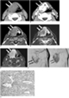
A precontrast CT scan revealed a 3 × 4 cm, well-defined, and slightly hypodense mass in the supraglottic larynx with preserved surrounding fat planes (Fig. 1A). In addition, the revealed the effacement of the aryepiglottic fold and obliteration of the pyriform sinus. Moreover, a marked heterogeneous enhancement was seen on a postcontrast scan (Fig. 1B). However, no evidence of adjacent cartilage destruction, extra-laryngeal extension, or cervical lymph node enlargement was observed. The MR imaging revealed that the mass was relatively hyperintense compared to the neck musculature on both the T1- and T2-weighted images (Fig. 1C, D). Further, a round-shaped region was observed at the posterior end of the tumor, which showed hypointensity relative to other regions of the tumor on both the T1- and T2-weighted images. The mass showed significant homogeneous enhancement after the administration of gadopentetate dimeglumine (Fig. 1E). The carotid angiograms showed a hypervascular tumor supplied by the bilateral superior thyroidal, bilateral lingual, and the left facial arteries with persistent central hyperstaining in the paramedian neck (Fig. 1F).
The preoperative radiologogic differential diagnosis was a hemangioma or possibly a hemangiopericytoma, neurogenic tumor, hypervascular sarcoma, or a goiter from ectopic thyroid tissue. The patient underwent a supraglottic partial laryngectomy. During surgery, a well encapsulated, round mass was found above the thyrohyoid membrane, as well as within the paraglottic and preepiglottic space. The mass was easily separated from the surrounding laryngeal tissue and completely excised. A pathological examination revealed a large well-demarcated fibrotic soft tissue mass. Under light microscopy, the tumor appeared as uniform spindle cells forming a solid pattern, with some areas showing prominent vascularity in a staghorn feature and hemangiopericytoma-like foci (Fig 1G). No evidence of mitosis or cellular atypism was observed. The round hypointense area on both the T1- and T2-weighted MR images corresponded to areas showing abundant deposits of collagen upon pathologic examination. The immunohistochemistry was positive for CD34 and vimentin (Fig. 1G); however, was negative for S-100 protein and keratin. Based on these features, the neoplasm was diagnosed as a benign SFT.
DISCUSSION
An SFT is an uncommon tumor that usually occurs in the pleura; however, it has also been reported in a number of extrapleural sites, such as the thoracic wall, mediastinum, pericardium, and abdominal cavity. Further, these lesions are rare in the head and neck; however, are occasionally encountered in the orbit, nasal cavity, paranasal sinus, nasopharynx, parapharyngeal space, thyroid gland, and larynx (1-6).
A review of previously reported cases (5-9) revealed that the average patient age at presentation of a laryngeal SFT is 52 years (29-73 years), with a male preponderance (male-female ratio of 5:1). All the documented cases of laryngeal SFTs were located, as in our case, in the supraglottic region. The major presenting symptoms were progressive hoarseness, foreign body sensation, cough, or even acute upper airway distress.
Eighty-seven percent of the reported SFTs have benign clinical behavior; therefore, a surgical resection is the treatment of choice and is curative in most cases (8). Previous reports have indicated that 12-35% of pleural SFTs have been associated with an invasive growth pattern or malignant histological features (9-11). However, SFTs of the head and neck generally have a benign course, with only a few malignant cases. To date, no cases of malignant laryngeal SFTs have been reported.
The diagnosis of an SFT depends mainly on its histologic appearance, usually after surgery. The SFT is mainly composed of spindle cells, which are randomly arranged in a collagenous background. Some cases have hemangiopericytoma-like areas with prominent branching vessels. The diagnosis of SFTs is also supported by an immunohistochemical profile. Tumor cells in SFTs are characteristically immunoreactive for CD34, CD99, and vimentin; however, they are usually negative for cytokeratin, S-100 protein, and smooth muscle actin (11).
In previous reports, SFTs displayed isoattenuation relative to the adjacent musculature on CT images, and showed enhancement following an intravenous contrast injection. The intratumoral low attenuation areas are correlated with myxoid or cystic degeneration (1-3). In our case, this low attenuation area is considered to be a slowly enhanced stromal area.
According to previous reports, the signal intensity of the SFT in our study was isointense relative to muscle on the T1-weighted MR images, and variable on the T2-weighted MR images. Further, it showed both heterogeneous and homogeneous enhancement. This variable signal intensity has been suggested to be caused by differences in the main components of the tumor, namely, the collagen and fibroblast content, as well as the presence of degeneration (3). Angiographic findings have rarely been described for cases of SFTs. In our case, an angiography revealed a hypervascular mass with persistent staining, which most likely reflected the abundant collagenous stroma of the tumor.
An interesting imaging feature in our case study was the large round area of hypointensity within the mass for both the T1- and T2-weighted MR images. Several previous MR studies also reported the linear or curvilinear hypointense areas within SFTs on T2-weighted images, and this feature was correlated with the hypocellular area, and more importantly, the collagenous sclerotic area (2, 3). This low signal intensity was larger and more round than those described in previously reported cases, and corresponded well to the abundant collagenous stroma for the pathologic correlation. Given the observations described in previously reported laryngeal SFTs, this large low signal intensity area on T1- and T2-weighted MR images may be a characteristic feature of laryngeal SFTs. Benign mesenchymal tumors of the larynx include hemangiomas, neural tumors, papillomas and a few other rare lesions. Cases of adult laryngeal hemangiomas are supraglottic, whereas those diagnosed in children are subglottic. Phleboliths are characteristic of hemangiomas; whereas, papillomas are mucosal tumors which typically affect the true vocal cords. However, little consideration is needed for these types of tumors in our case. Neurogenic tumors originate from the superior laryngeal nerve, and may secondarily involve the arytenoid muscles and the aryepiglottic folds. Other rare lesions include granular cell tumors, leiomyomas, and rhabdomyomas (12). However, these submucosal tumors of the larynx show non-specific radiologic findings.
We have presented a case of an SFT in the submucosal region of the larynx and reviewed its imaging features. As seen in this case report, the imaging features for SFTs of the submucosal region of the larynx are nonspecific, and are similar to those of other more common tumors such as hemangiopericytomas, neurogenic tumors, and soft tissue sarcomas. Consequently, when a well-marginated mass involving the larynx shows intense contrast enhancement on both CT and MR, heterogeneous and large round hypointensity on T2-weighted MR images, and hypervascularity with persistent staining on an angiography, an SFT should be included as part of the differential diagnoses.
 XML Download