

 PDF
PDF ePub
ePub Citation
 Print
Print


Although the pathogenesis of focal nodular hyperplasia (FNH) is not well understood, the general consensus is that hepatic vascular abnormality plays an important role in its development (1, 2). Herein we present a rare case of multiple FNH that was associated with portal vein atresia and portopulmonary hypertension in a young female patient.
CASE REPORT
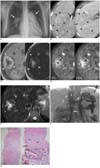
A 30-year-old woman was admitted to our hospital to further evaluate her hepatic nodules. She had experienced dyspnea on exertion since the time of attending middle school. She had been diagnosed as having pulmonary hypertension and multiple liver nodules ten years earlier. She has been treated with diuretics, calcium channel blockers and sildenafil. She had experienced transient aggravation of her dyspnea one week before her recent delivery. Her platelet count was 99,000/mm3. Blood chemistry tests showed normal levels of transaminases, alkaline phosphatase and bilirubin. The serologic markers for hepatitis B and C virus were negative. Chest radiography (Fig. 1A) showed enlarged pulmonary arteries and cardiomegaly.
Ultrasonography showed numerous hyperechoic and isoechoic nodules of various sizes in the liver. On CT, the liver showed heterogeneous contrast enhancement due to the numerous nodules (Fig. 1B). These nodules were heterogeneously hyperattenuating during hepatic arterial phase, and they showed contrast material washout during the portal venous phase. Persistent central scar-like enhancement was found in some of the nodules. The hepatic arteries were hypertrophied. The portal vein was not visualized; however, splenic varix and splenorenal shunt were present.
The hepatic nodules had high signal intensity with central portions of low signal intensity on the T1 weighted magnetic resonance images, and the hepatic nodules were of low signal intensity with central portions of high signal intensity on the T2 weighted images (Fig. 1C). On the gadolinium enhanced dynamic MR scans, the central hypointense portions of the nodules were filled with contrast media for a length of time and they showed persistent intense enhancement (Fig. 1D). On the gradient echo sequence images obtained after superparamagnetic iron oxide enhancement, the nodules showed a drop in signal intensity except at their central portions (Fig. 1E).
The patient underwent percutaneous ultrasound-guided needle biopsy. The biopsied tissue core demonstrated a dense fibrous septum containing numerous vascular structures at low power (Fig. 1G). Higher power magnification of the portal tracts revealed thick-walled vessels, and some had eccentric thickening. The surrounding hepatic parenchyma had slightly increased cellularity, with increased liver cell plate thickness. These findings were consistent with FNH.
Additional abdominal angiographies were obtained during the diagnostic cardiography to evaluate her cardiac function. Hepatic arteriography showed numerous stained nodules in the liver with hypertrophied hepatic arteries. The delayed phase of the superior mesenteric arteriography showed the absence of the main portal vein flow, and the portosystemic collaterals drained into the inferior vena cava (Fig. 1F).
DISCUSSION
In this case, we observed the coexistence of multiple FNH, portal vein atresia and portopulmonary hypertension. There have been several case reports describing similar combinations of two or three (3) of these abnormalities.
Multiple FNH syndrome has been described and this refers to multiple FNH in combination with liver hemangiomas and vascular malformations, including hepatic arterial dysplasia, portal vein atresia and aneurysms of the brain, or pulmonary arterial hypertension and/or intracranial tumors (1).
For the association between multiple FNH and portal vein atresia (1, 4-7), it has been postulated the latter can play a role in the development of the former since a decreased portal flow can alter the delivery of hepatotrophic factors that affect the hepatic regenerative capacity, while the increased compensatory hepatic arterial flow can also contribute to the development of nodular hepatocellular lesions such as FNH, regenerative nodular hyperplasia and large non-cirrhotic regenerative nodules (8-10).
For the association between portal vein atresia and pulmonary hypertension, it has been suggested that vasoactive substances bypassing the liver through portosystemic collaterals can cause pulmonary arterial spasm and thrombosis, which is a condition called portopulmonary hypertension (11). The main clinical presentation of portopulmonary hypertension is hemodynamic failure and the severity of portopulmonary hypertension may not parallel the severity of liver failure; this is different from hepatopulmonary syndrome, which is another pulmonary vascular disorder that occurs with portal hypertension (12).
For the association between FNH and pulmonary hypertension, it has been suggested that chronic congestion of the hepatic sinusoids can prolong the exposure of the liver to blood-borne hepatotrophic substances that induce a hyperplastic response of the hepatic parenchyma, and this stimulates the growth of nodular hepatocellular lesions such as FNH, which is a compensatory vicious cycle (3).
In our case, the chronological or causative relationships are not clear between the multiple FNH, portal vein atresia and portopulmonary hypertension. Her history of dyspnea since her teenage years suggests the portal vain atresia could be congenital or it had occurred in young childhood. Nevertheless, it is plausible that the portal vein atresia and the resultant portopulmonary hypertension played major roles in the development of the multiple FNH in this case.
In conclusion, we present radiological findings of a rare case of multiple FNH associated with portal vein atresia and portopulmonary hypertension. This case illustrates and supports the pathophysiologic hypotheses that have previously proposed for the concurrent existence of these three abnormalities.
 XML Download