

 PDF
PDF ePub
ePub Citation
 Print
Print


An angiomyofibroblastoma (AMFB) is a relatively recently described tumor in the expanding spectrum of benign mesenchymal tumors that are found in the female lower genital tract. Fletcher and colleagues (1) first described AMFB in 1992. The importance of this rare neoplasm is related to its potential mimicry of more infiltrative and prognostically less favorable lesions, such as aggressive angiomyxomas, that are found in the same anatomic area (2).
We present a case of AMFB that arose from the posterior perivesical space, with an emphasis of the magnetic resonance (MR) imaging features of the disease.
CASE REPORT
A 48-year-old woman presented with a vaginal mass that was an incidentally detected lesion during gynecological cancer screening. A physical examination revealed a hard mass on the anterior wall of the vagina, and the laboratory results were unremarkable.
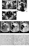
MR imaging was performed on a Gyroscan Intera 1.5 T unit (Philips, Best, The Netherlands). Axial and coronal T1-weighted images (TR 739/TE 25) with a 5 mm slice thickness as well as axial and coronal T2-weighted images (TR 4218/TE 100) with a 5 mm slice thickness were obtained. In addition, axial and sagittal gadolinium-enhanced T1-weighted images (TR 139/TE 25) with a 5 mm slice thickness as well as sagittal dynamic gadolinium-enhanced T1-weighted images (TR 165/TE 4.6) with an 8 mm slice thickness were obtained. The mass was demonstrated as a well-defined, oval-shaped mass in the posterior perivesical space that measured 38 × 35 × 28 mm. This mass was located posterior to the inferior wall of the bladder and the upper portion of urethra and compressed both structures. Furthermore, the mass was located anterior to the vagina and uterine cervix and was separated from these structures (Fig. 1). The mass displayed heterogeneous intermediate signal intensity with focal nodular or curvilinear dark signal intensity areas within the tumor as seen on T2-weighted images (Figs. 1A, C). On T1-weighted images, the signal intensity of the mass was similar to that of skeletal muscle (Fig. 1B). After intravenous injection of gadopentetate dimeglumine (Magnevist, Schering, Erlangen, Germany), the mass showed strong and homogeneous enhancement on T1-weighted images. On contrast-enhanced dynamic MR images obtained at 30-seconds, 1-, 2-, and 3-minutes after the administration of contrast material, the mass showed fast and persistent enhancement on early and late phase images (Figs. 1D-F). The preoperative diagnosis based on these MR findings included a leiomyoma from the urinary bladder or urethra, a neurilemmoma, and a soft tissue sarcoma.
At surgery, a well encapsulated, round, hard mass was found that arose in the posterior perivesical space. The mass was easily separated from the adjacent organs such as the urinary bladder, urethra, vagina, and uterine cervix. The mass was completely excised. A histological examination of the tumor specimen showed monotonous small round or ovoid stromal cells around the vessels without spindle cell changes. The tumor was characterized by the presence of numerous small- to medium-sized and thin-walled intratumoral blood vessels. A variable number of inflammatory cells and myxoid fibrous stroma were also detected in the lesion. From immunohistochemical analysis, the tumor cells were positive for actin (Figs. 1G, H). These distinctive histological features were compatible with a diagnosis of an AMFB.
DISCUSSION
Angiomyofibroblastoma predominantly occurs in middle-aged premenopausal women and involves the genital region (1-6). The most common site is the vulva, followed by the labia major, vagina, periclitoris and perineum (3). Since an AMFB has a benign clinical behavior, it should be differentiated from an aggressive angiomyxoma, cellular angiofibroma, and other myxoid tumors of the genital area where radical surgical treatment is indicated (4). To the best of our knowledge, this is the first reported case in the literature of an AMFB that arose in the posterior perivesical space.
Histologically, an AMFB is a benign soft tissue tumor of myofibroblastic differentiation and represents neoplastic proliferation of stromal cells (1). An AMFB is distinguished from an aggressive angiomyxoma by its higher cellularity, by the frequent presence of plump stromal cells, and to a lesser degree with stromal myxoid changes (5).
To date, only two previous reports have described imaging features of this rare neoplasm. The first reported case was a 24-year-old woman that presented with a round mass that extended to the paraurethral region with homogeneous intermediate signal intensity as seen on a T2-weighted MR image (3). The second reported case was a 46-year-old woman that presented with a perineal mass that was mainly well delineated and hyperintense, similar to the ischiorectal fat on a T2-weighted MR image, and homogeneously enhanced on a contrast-enhanced MR image (6).
The MR findings in our case appear to be quite different from those in previous reports, except for the well-defined margin of the tumor. In our case, the T2-weighted MR images of the tumor showed heterogeneous intermediate signal intensity. We also observed nodular or curvilinear dark signal intensities within the tumor on T2-weighted MR images. We consider this finding to be of interest. The dark signal intensities corresponded well to the areas of hypocellularity and abundant collagenous stroma as seen on the pathological specimen.
Another important radiological finding is the strong and homogeneous enhancement of the tumor (6). This characteristic most likely reflects prominent vascularity of a tumor of myofibroblastic differentiation. In contrast-enhanced dynamic MR images, the mass in our case showed a similar enhancement pattern to the uterus, which was a strong and homogeneous enhancement at early phase and persistent enhancement at late phase. This marked delayed contrast enhancement may be due to a prolonged high concentration of contrast medium in the tumor tissue, caused by delayed washout of contrast medium from the abundant fibrous tissue.
In summary, we have presented a case of AMFB in the posterior perivesical space and have reviewed the MR imaging features. As seen in this case and in previously reported cases in the literature (3, 6), the MR features for AMFB are nonspecific and are similar to the features of the other more common pelvic tumors such as leiomyomas, neurogenic tumors, soft tissue sarcomas, or fibromyxoids or angiomyxoid mesenchymal neoplasms. However, where a well-marginated mass that involves the external genitalia in women shows heterogeneous hypointensity on T2-weighted MR images and intense and delayed contrast enhancement, an AMFB should be included in the differential diagnosis.
 XML Download