

 PDF
PDF ePub
ePub Citation
 Print
Print


Atypical pattern of transient left ventricular contractile dysfunction confined to the apical and mid-ventricular portions of the heart after physical or emotional stress, which mimics acute myocardial infarction but shows no angiographic evidence of obstructive epicardial coronary artery disease, has been reported on and it's been named 'stress-induced cardiomyopathy' (1) or 'transient left ventricular apical ballooning' (2). The mechanism for this malady remains unclear, although exaggerated sympathetic stimulation has been considered to play a major role (3, 4). From a diagnostic standpoint, magnetic resonance imaging (MRI) has been reported in a few studies to be able to provide additional information to exclude myocardial infarction at the level of the myocardium by demonstrating the absence of delayed hyperenhancement (3, 5). In this case report, we suggest that MRI might be an integrated imaging diagnostic tool for the diagnosis of this syndrome, which demonstrated characteristic apical contractile dysfunction with performing cine MRI, the absence of significant coronary artery stenosis with performing coronary MR angiography and the absence of myocardial infarction with performing contrast-enhanced delayed MRI.
CASE REPORT
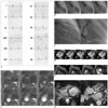
A 66-year-old woman presented to the emergency department with chest pain, dyspnea and sweating for the previous 4 hours after making a speech in front of a large group of people. She had been taking medicine for hypertension. The initial electrocardiogram on admission showed T-wave inversion in leads V3 through V6, and Q-wave in leads V4 through V6 (Fig. 1A), and this was all newly developed compared to the ECG taken one year previously. The troponine-T, CK-MB and CK levels were slightly elevated to 0.565 ng/mL (normal range: 0-0.1 ng/mL), 10.98 ng/mL (normal range: 0-5 ng/mL), and 164 IU/L (normal range: 32-135 IU/L), respectively. An emergency echocardiography showed a 49% ejection fraction and extensive akinesis from the mid to apical left ventricle, except for the base. An aortogram showed the same findings as echocardiography, that is, akinesis from the mid to apical left ventricle with sparing of the basal segments (Fig. 1B). No intraventricular pressure gradient was observed. However, a coronary angiography showed no significant stenosis of the epicardial coronary arteries (Fig. 1C). Four days later, she underwent MRI on a 1.5-T unit. Cine imaging, the contrast-enhanced first-pass and delayed imaging, and the respiratory compensated coronary angiography with a navigator echo were performed. There was severe hypokinesis from the mid to apical left ventricle with sparing of the basal segments (Fig. 1D). The dysfunction of the left ventricle was slightly improved compared to the initial left ventriculogram. Mild myocardial wall thinning at the apical segments was observed on cine MRI. There was no perfusion abnormality (Fig. 1E) or hyperenhancement of the myocardium (Fig. 1F). Coronary MR angiography showed no significant stenosis of the major epicardial coronary arteries (Fig. 1G). The plasma catecholamine levels were not checked. She completely recovered her ventricular function and from her symptoms after two weeks, the former as determined on follow-up with performing echocardiography, with only supportive treatment.
DISCUSSION
A report from Japan demonstrated that incidence of acute cardiovascular events after an earthquake was triple the incidence before the earthquake (6). This phenomenon has recently come to be recognized as a reality in clinical medicine and it is reported on as a novel cardiac syndrome. Dote et al. first described this unique syndrome in 1991 in five patients who presented with acute chest pain and electrocardiographic abnormalities that matched the manifestation of acute myocardial infarction, but they had no coronary artery stenosis (7). Since then, this syndrome has been reported, mostly from Japan, by the name of 'tako-tsubo cardiomyopathy' or 'ampulla cardiomyopathy', which reflected the characteristic shape of the left ventriculogram at end-systole that resembled a Japanese pot (tako-tsubo) with a narrow neck and wide base. The tako-tsubo-like shape results from apical and mid-ventricular akinesis with the basal hyperkinesis, which is not easily explained by any distribution of coronary artery disease; this has been considered as one of the evidences that this syndrome is a distinct entity that differs from acute myocardial infarction. Since this characteristic left ventricular dysfunction resolved in days or weeks for most reported cases, 'transient left ventricular apical ballooning' has recently been used to name this syndrome and to emphasize the reversibility of the left ventricular dysfunction.
The reversible ventricular dysfunction seen in this syndrome can be attributed to myocardial stunning, which is defined as prolonged postischemic left ventricular dysfunction after a short duration of myocardial ischemia. Myocardial stunning in the syndrome is precipitated by either intense emotional or physical stress. Markedly elevated and sustained levels of plasma catecholamines and stress-related neuropeptides that are related to intense stress in most patients with the syndrome have been reported, and they are considered to play a central role between the severe stress and the myocardial stunning in this syndrome (3, 4). However, the mechanism of how catecholamine and stress-related neuroepeptides can induce myocardial stunning is not fully understood. The possible mechanisms that have been suggested to account for the stress-related myocardial stunning include ischemia resulting from vasoconstriction of the coronary arteries that's due to increased sympathetic tone, sympathetically-mediated microcirculatory dysfunction and catecholamine-induced direct myocyte injury. Contraction-band necrosis, which is compatible with catecholamine-induced myocyte damage, was found on myocardial biopsy in some patients with this syndrome (3). The strong female predominance of this syndrome is not understood either.
The universally accepted diagnostic criteria for transient left ventricular apical ballooning are not yet available. A report from the Mayo Clinic suggested the diagnostic criteria for the syndrome, which should meet all of the following; 1) transient akinesis or dyskinesis of the left ventricular apical and mid-ventricular segments, 2) absence of obstructive coronary artery disease or acute plaque rupture, 3) new electrocardiographic abnormalities, and 4) exclusion of any apparent etiology causing acute myocardial stunning, including head trauma, intracranial or subarachnoid bleeding, pheochromocytoma, obstructive epicardial coronary artery disease, myocarditis and hypertrophic cardiomyopathy (8). Coronary arterial imaging is a mandatory procedure to determine these criteria. Even though coronary arterial imaging can reveal no obstructive epicardial coronary artery disease, syndrome X or idiopathic dilated cardiomyopathy should be excluded from the differential diagnosis; however, transient dysfunction of the left ventricular apical and midventricular segments is not a typical presentation for these entities. A prior stressful event as a precipitating factor or increased catecholamines levels are not a diagnostic requirement, although both are helpful to diagnose the syndrome. In our case, the characteristic tako-tsubo-like left ventricular geometry was demonstrated by cine MRI, like was demonstrated by left ventriculography and echocardiography. The absence of wall thinning was additional information provided by MRI. The absence of obstructive epicardial coronary artery disease was confirmed by respiratory compensated coronary MR angiography, the same was done by X-ray coronary angiography. In addition, contrast-enhanced delayed MRI revealed an absence of hyperenhanced myocardial segments, which implicated the absence of acute or chronic myocardial infarction, regardless of coronary artery disease (9). Although we did not performed stress perfusion MRI in the patient, the lack of hypoperfusion on the first-pass imaging at rest suggested there was neither profound compromise of the epicardial flow nor any microvascular obstruction. Therefore, MRI may not only substitute for X-ray coronary angiography and left ventriculography, but it can also confirm that acute or chronic myocardial infarction is absent and the left ventricular dysfunction is reversible (9). The morbidity and mortality related to invasive X-ray coronary angiography can be avoided by using MRI for the diagnosis of this syndrome.
Although the mechanism of transient left ventricular apical ballooning syndrome has still not been fully elucidated, a typical presentation of tako-tsubo-like apical ballooning, the absence of obstructive epicardial coronary artery disease and the reversibility of myocardial dysfunction are very characteristic and these factors can be determined by performing MRI.
 XML Download