

 PDF
PDF ePub
ePub Citation
 Print
Print


Mesenchymal chondrosarcoma is a rare and more aggressive variant of conventional chondrosarcoma. It represents approximately 1% of all chondrosarcomas and it carries a poor prognosis. It can occur in extraskeletal locations and mainly in the soft tissues of the orbit, the cranial and spinal meningeal coverings and the lower limbs (1, 2). To the best of our knowledge, there has been no reported case of primary extraskeletal mesenchymal chondrosarcoma of the pancreas. Only two instances of metastatic chondrosarcomas in the pancreas have been reported in the literature (3, 4). We report here on a case of primary mesenchymal chondrosarcoma arising from the pancreas in a 41-year-old man.
CASE REPORT
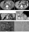
A 41-year-old man presented with a 1-month history of abdominal pain. His previous medical and familial histories were unremarkable. Unenhanced transverse CT scan of the abdomen revealed an ill defined, lobulated mass that measured about 13 × 12 × 7 cm in size and it appeared as a heterogeneously low attenuated mass with numerous areas of coarse calcification. It was located in the left side of the retroperitoneal space and involved the body and tail of the pancreas (Fig. 1A). Contrast-enhanced CT was performed after mechanical injection of 130 cc of nonionic iopromide (300 mg/ml of iodine) into the antecubital vein at a rate of 2.65 ml/sec. After initiating infusion of contrast material, we used scanning delays of 30 seconds before starting to obtain the arterial phase images. Imaging in the portal venous phase was performed after a scanning delay of 70 seconds from the initiation of infusion of contrast material. On the contrast-enhanced transverse CT scan obtained during the portal venous phase, subtle enhancement was noted in only the periphery of the mass and most of the mass remained without enhancement (Fig. 1B). The contrast-enhanced coronal reformatted image showed the ill defined necrotic low attenuated mass with obliteration of the splenic vein by the mass, and there was the development of multiple collateral veins (Fig. 1C). Portal venography via the celiac axis also showed tumor invasion to the splenic vein (Fig. 1D). The patient underwent exploratory laparotomy and mass excision was done. The specimen was revealed to be a large, firm, fixed hypervascular mass arising from the pancreas, and it occupied the left upper quadrant of the abdomen. A large multilobulated mass that measured 15 cm in maximum diameter was located in the body and tail of the pancreas, and it was attached to the spleen and transverse colon (Fig. 1E). Microscopically, the chondroid zone was surrounded by proliferation of undifferentiated cells with abrupt transition (Fig. 1F). The pathological diagnosis was extraskeletal mesenchymal chondrosarcoma arising from the pancreas with invasion to the splenic vein.
DISCUSSION
Mesenchymal chondrosarcoma was first reported by Lichtenstein and Bernstein in 1959 (5) and this type of tumor discovered in the soft tissue by Dowling in 1964 (6). It is an uncommon tumor of the bone and soft tissues, and is defined as the coexistence of nests of well-defined cartilaginous tissue within a proliferation of primitive mesenchymal cells. Mesenchymal chondrosarcoma is a malignant neoplasm with high metastatic potential, particularly to the lungs, and it tends to occur preferentially in bones and soft tissues (1). In contrast to conventional chondrosarcoma for which only 1% of tumors are extraskeletal, mesenchymal chondrosarcoma developed in the soft tissues in almost half the reported cases (2). Extraskeletal mesenchymal chondrosarcomas are located mainly in the soft tissues of the orbit, the cranial and spinal meningeal coverings, and the lower limbs, and particularly the thigh. In the rare instances, this type of tumor has been found in the mediastinum, hand musculature, retroperitoneum and kidney (1). Extraskeletal mesenchymal chondrosarcoma affects women slightly more often than men, whereas men are predominate for skeletal and extraskeletal conventional chondrosarcoma (7). Extraskeletal mesenchymal chondrosarcoma has two peak age incidences in adults, depending on its location: at 23.5 years for central nervous system involvement and at 43.9 years for soft-tissue and/or muscular involvement (8). Mesenchymal chondrosarcoma, regardless of its origin, often displays a lethal clinical course with a 5-year survival rate of 55% and a 10-year survival rate of 27% (5); radical excision of the tumor is the recommended treatment (7).
Conventional radiography, CT and magnetic resonance of extraskeletal mesenchymal chondrosarcoma demonstrate several overlapping features with those of conventional chondrosarcoma (9). Conventional radiography and CT depict extraskeletal mesenchymal chondrosarcoma as a soft-tissue mass with ring, arc, stippled and highly opaque calcifications. Contrast-enhanced CT depicts a soft tissue mass that consists of an enhancing viable tumor portion in the periphery and central focal low attenuation areas that possibly represent necrosis (2). Our patient showed similar CT findings. Extraskeletal mesenchymal chondrosarcoma also shares imaging findings with other skeletal and soft-tissue tumors. Chondroid-type calcifications and lobulation are features of both benign and malignant cartilaginous tumors, although these tumors are not usually found in extraskeletal locations (2). Their gross appearance is often large (nearly 10 cm at the greatest dimension), lobulated and well circumscribed, and they contain cartilaginous foci. The histologic diagnosis is definitive, showing a combination of highly cellular, undifferentiated mesenchymal cells and islands of well-differentiated cartilage (9). Histologically it is defined by the coexistence of nests of well-defined cartilaginous tissue and calcification within a proliferation of primitive or undifferentiated mesenchymal cells (1).
Although the imaging findings of lobulated, enhancing masses with cartilaginous calcifications on plain radiographs and CT scans are not specific for extraskeletal mesenchymal chondrosarcoma, they could reflect the unique histopathologic characteristics of vascular undifferentiated mesenchyme that surrounds foci of differentiated cartilage (2).
In summary, we present here a case of primary extraskeletal mesenchymal chondrosarcoma that arose from the pancreas. Radiologically, it manifested as a necrotic soft tissue mass with chondroid calcifications.
 XML Download