

 PDF
PDF ePub
ePub Citation
 Print
Print


There has been debate concerning the relative contribution of anatomic narrowing versus increased airway compliance in the development of obstructive sleep apnea (OSA) (1-15). Dynamic imaging studies have proven helpful in the evaluation of these relationships as they can be used to evaluate both anatomy and dynamic motion of structures (16-27). Though theories incorporating both anatomic obstruction and airway compliance have been proposed (1-15), the relative importance of anatomic obstruction and airway compliance has been debated (12, 13).
The purpose of this study is to evaluate the distensibility and collapsibility of the upper airway as depicted on cine MRI in children with tracheotomy tubes that were evaluated for anatomic reasons of decannulation difficulty. Cine MR images were reviewed for the variation in volume of the upper airway over time, in a comparison of whether the tracheotomy tubes were either uncapped or capped. The assumption is that with the tracheotomy tube is capped, patients breathe through the supra-glottic airway and have greater pressure changes over time in the airway, as compared to when they breathe through the tracheotomy tube and there is no airflow through the supraglottic airway. This follows the Bernoulli principle, which states that the inward, collapsing pressure exerted on the airway is proportional to the velocity of air flowing through the airway. Through a comparison of airway motion at two different pressure states (uncapped and capped), we can speculate on the role of compliance in the pathogenesis of airway obstruction in children.
MATERIALS AND METHODS
Inclusion criteria in this study consisted of patients referred for cine MRI to evaluate potential supra-glottic causes of the inability to tolerate tracheotomy tube decannulation. There were seven patients enrolled in the study. The primary clinical problem encountered in these patients preventing decannulation was OSA. Diagnoses of each patient were recorded. There were six boys and one girl, with a mean age of eight years (range: 2 to 15 years). Cine MR images were reviewed for the variation in the volume of the upper airway over time in a comparison when the tracheotomy tubes were uncapped and capped.
Our institutional review board approved the review of the patient records. As this was a retrospective review of clinical data, our institutional review board waived informed consent. The reviewed data was de-identified and stored in a secure database.
MR Imaging
All of the MRI examinations were performed on sedated patients. No patients had complications because of sedation. The clinical protocol for obtaining MR images to evaluate the supra-glottic airway included multiple sequences: T1-weighted spin echo sagittal and axial images, fast spin echo inversion recovery T2-weighted sagittal and axial images, and MR cine images in the sagittal midline and axial planes at the level of the base of the tongue. Examinations were performed with a 1.5T MRI scanner with the use of a head-neck vascular coil. The patients were imaged with the neck in a neutral position while supine. The airway was imaged from the most superior aspect to the level of the lower cervical trachea.
For the purposes of the quantitative image analysis for this study, only the sagittal cine MR images were evaluated. Our current process does not allow the evaluation of both sagittal and axial data sets simultaneously, and the sagittal plane was chosen as it was considered as more representative of the entire supraglottic airway. Cine MRI sequences were generated by a fast gradient echo series with the following technical parameters: 8.2/3.6 (repetition time msec/echo time msec), an 80˚ flip angle, and an 8 mm slice thickness. One hundred twenty-eight consecutive cine MR images were obtained at the same midline sagittal location in approximately 2 minutes. The midline sagittal plane was determined from the localizer images and the static transverse and sagittal MR images. These images were obtained in the midline sagittal plane and in the transverse plane at the base of the tongue and displayed in a cine format to create a real-time "movie" of airway motion. Axial and sagittal MR cine images were each obtained twice: once with the tracheotomy tube uncapped and the subject breathing through the tracheotomy tube, and once with the tracheotomy tube capped and the subject breathing via the native airway. The MR images were reviewed by one observer for evidence of anatomical abnormalities that may lead to occlusion of the upper airway.
Volume Segmentation of the MR Data
Volume segmentation allows objective and precise quantification of airway size and it can be automated to analyze large numbers of images in a consistent manner. The midline sagittal cine MR images obtained with the tracheotomy tube capped and uncapped underwent volume segmentation of the nasopharynx and the hypopharynx to evaluate changes in the airway volume over time. Cine MR images, in the Digital Imaging and Communications in Medicine (DICOM) file format, were transferred to a computer workstation. An in-house IDL (Research Systems, Inc, Boulder, CO) based image analysis software program was used to conduct volume segmentation of these images. The area of a single slice MR image was converted to volume by multiplying by the slice thickness. The entire cine MRI time course of the 128 separate images was converted into a single data matrix of signal intensity.
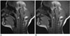
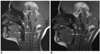
A region of interest for volume segmentation was manually selected to evaluate either the nasopharynx or the hypopharynx (Fig. 1). The nasopharynx was defined antero-superiorly by the sphenoid and posterior choanae, posteriorly by the anterior aspect of the cervical vertebrae and the adenoid tonsils, and inferiorly by the soft palate and hard palate. The hypopharynx was defined superiorly by the base of the adenoid tonsils, posteriorly by the posterior pharyngeal wall, inferiorly by a horizontal plane at the level of the hyoid bone, and anteriorly by the posterior aspect of the tongue (Fig. 1). The matrix of intensity data within the region of interest was then analyzed using a k-means clustering algorithm that segments the region of interest using signal intensity for the entire data set (all slices over all time). Three segments of intensity were selected as this yields the most accurate depiction of airway volume as this best represented the anatomy of the patent airway regions (26). The image segment representing patent airway intensity was selected and the airway volume was plotted as a function of time. Numerical characterization of the airway size allowed statistical analysis of airway dynamics and objective comparison of uncapped and capped images.
The airway volume, standard deviation, and the range of airway volumes were calculated for both uncapped and capped images for each individual. The mean airway volume was calculated by averaging the individual airway volumes measured for each image of the cine MR imaging sequence. The change in volume over time can be normalized by use of the average airway volume for the subject and can be plotted across the time course (change in volume divided by mean volume). Normalization allows for the change in volume of the area to be viewed in relationship to the relative size of the subject's airway, as this may vary between subjects of different sizes, ages and the imaging plane. The normalized range and standard deviation for the normalized range (coefficient of variance) was calculated. These parameters are measures of individual airway dynamics.
Statistical Analysis
Differences in the mean airway volume, airway volume standard deviation, range of airway volumes, coefficient of variance, and normalized range between capped and uncapped tubes for both the nasopharynx and the hypopharynx were compared for statistical significance and differences using the signed rank test. Statistical significance was defined as p < 0.05. Analyses were performed using SAS Statistical Software, Version 8.2 (SAS Institute, Cary, NC).
RESULTS
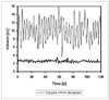
The results compiled from volume segmentation of cine MRI comparing volume changes in patients with the tracheotomy tubes both capped and uncapped are summarized in Table 1, and are illustrated in Figures 2-4. There were statistically significant differences seen in the nasopharynx between the uncapped and capped tubes for the mean airway volume, the airway volume standard deviation, and the airway volume range. There were statistically significant differences in the hypopharynx between the uncapped and capped tubes for the airway volume standard deviation and the airway volume range, but not for the mean airway volume. The mean airway volume, the airway volume standard deviation, and the airway volume range of both the nasopharynx and hypopharynx were larger in the capped tubes. The increase in airway dynamics with tracheotomy tube capped is demonstrated in Figure 3.
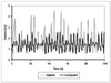
Variability attributed to airway size was isolated from airway dynamics by normalizing the airway volume by the average airway volume of the patients. The normalized airway standard deviation (coefficient of variance) and the normalized airway volume range were not statistically significant for the nasopharynx, but were statistically significant for the hypopharynx (Fig. 4).
DISCUSSION
The relative importance of the roles of anatomic obstruction and airway compliance in the development of OSA has been subject to debate (12, 13). The role of airway compliance has been addressed in the literature, and more focus has been placed on the functional aspects of OSA (1-15). During sleep, there is decreased neural output to the upper airway dilators, which leads to decreased muscular tone (11). This decreased muscular tone results in pharyngeal narrowing, increasing airflow velocity through this narrowed segment (11). The increased velocity further reduces intraluminal pressures (or increases the inward, collapsing pressures), promoting further pharyngeal narrowing and eventual collapse (11). This effect is known as Bernoulli's Principle (11). The principle states that the inward pressure on a tube is proportional to the velocity of the fluid flowing through the tube. This relationship is described in the following equation:
P = ρv2/2
where P is the inward pressure, ρ is the density of the fluid, and v is the velocity of the fluid flowing through the tube. It has been reported that patients with upper airway obstruction have narrowed pharyngeal segments (10, 14, 15, 28, 29). As the velocity of air in these narrowed segments increases, Bernoulli's principle demands that the inward pressure in these segments increases as well. This has an additive effect on an already narrowed airway, which increases the possibility of collapse.
Patients with difficulty in decanulation of tracheotomy tubes may have anatomic or elastic issues predisposing them to airway obstruction. These abnormalities may be depicted on MRI cine images with the tracheotomy tubes capped. These children with tracheotomy tubes provide us with the ability to image the airway at two different states of airway dynamics: with and without a velocity of air traveling through the supraglottic airway. The statistically significant differences in airway motion parameters show that Bernoulli's Principle does apply in the human airway. Those patients who have increased compliance of the upper airway would be even more susceptible to the above sequence of events. This would lead to increased collapsibility and greater severity of disease, regardless of the anatomical factors present.
We have presented a series of seven patients who have been difficult to decannulate from the tracheotomy tubes manifested as OSA.
When these tracheotomy tubes were capped, the upper airway showed significant variability in the airway volume standard deviations, and airway volume ranges. This quantitative variability corresponds to the upper airway distending and collapsing over time, and may support the theory that increased compliance of the upper airway has a significant role in OSA in this specific group of patients.
After the normalization of data, the variability of the nasopharynx was found to be not statistically significant. This may not be unexpected, as the nasopharynx is much smaller and has less volume for variability as compared to the hypopharynx. Thus, the variability we observed in the nasopharynx under the two pressure states was not significant, and most likely was secondary to the size of the nasopharynx.
Whereas all comparisons of the variance of airway volume over time were significant for the hypopharynx, including normalized measures of airway variance, the mean airway volume of the hypopharynx was not statistically significant when comparing uncapped with capped tubes as determined from the cine MR images. This finding does not infer that the capped airway was static; in fact, the significance of the airway volume standard deviation and the airway volume range of the hypopharynx provide evidence that the airway was not static, but rather distending and collapsing, as demonstrated in Figure 4. The hypopharynx was found to be significantly more variable in the capped tubes as seen on the cine MR images, but just with a similar average volume as compared with the uncapped tubes as seen on the cine MR images. In addition, findings in these patients with difficulty in tracheotomy tube decannulation that was manifested as OSA may not be applicable to other patients with OSA.
This study does have some limitations, including a small number of patients that were evaluated. In addition, many of these subjects had supraglottic anatomic abnormalities. These anatomic abnormalities most likely contributed to the airway dynamics seen. Therefore, airway compliance issues were not the only issues effecting airway dynamics.
In conclusion, there is a significant change in airway dynamics in children with tracheotomy tubes when breathing via the upper airway as compared to breathing via the tracheotomy tube and thus at two different states of airway flow. This demonstrates that airway dynamics are significantly different at those two different states of airway flow and that Bernoulli's Principle applies to the human airway. These findings further raise the question of airway compliance may play a significant role in the development of OSA.

 XML Download