

 PDF
PDF ePub
ePub Citation
 Print
Print


Amesenchymal hamartoma of the liver is an uncommon benign tumor mostly occurring in children, primarily in the first two years of life. It is thought to be a developmental anomaly of the portal connective tissue rather than a true neoplasm, and is composed of both mesenchymal and epithelial components (1).
Most of the reported cases of a hepatic mesenchymal hamartoma have described a large multiloculated mass with variable septa and cystic spaces (1-5). Rarely, a predominantly solid form of a mesenchymal hamartoma containing a larger amount of hepatocytes has been reported in the literature (6-9).
Although several reports have described the radiological findings of a mesenchymal hamartoma of the liver (3-6), little is known about the spectrum of radiological findings in a series of cases. The aim of this study was to describe a spectrum of the radiological findings of a mesenchymal hamartoma of the liver in children.
MATERIALS AND METHODS
Subjects
We retrospectively reviewed the radiological and clinical findings of 13 patients with a pathologically confirmed mesenchymal hamartoma of the liver. The institutional review board approved the review of the radiological and clinical data for this study and waived the requirement for informed consent. A computed hospital information system was used to identify all patients with a pathologically proved mesenchymal hamartoma during a 15-year period. The pathological diagnosis for a mesenchymal hamartoma was based on overgrowth of the mesenchymal stroma and the proliferation of architecturally abnormal bile ducts with or without a cystic change, accompanied by periductal collaring of the stromal cells (1, 9).
Imaging and Analysis
Three pediatric radiologists retrospectively reviewed the radiological features of the hepatic mesenchymal hamartomas by consensus.
CT scans were obtained with various third generation CT scanners at 80-120 mA, 100-120 kVp, and 5- to 10-mm thick sections. CT scans were obtained after an intravenous bolus injection of contrast media and portal venous phase scans of the liver were available for all patients. Pre-contrast CT scans were available for ten patients.
CT images of the tumor were evaluated for location, size, presence of septa within the cystic portion, CT attenuation of the tumor relative to the surrounding hepatic parenchyma (low, iso, or high), pattern of contrast-enhancement (homogeneous or heterogeneous enhancement of the solid portion, and septal enhancement of the cystic portion) and presence of calcification on a pre-contrast CT scan. According to the extent of cystic component on the CT scans, we classified each tumor into one of three types: 1) multiseptated cystic - a purely cystic tumor with multiple thin septa; 2) mixed cystic and solid-a partially cystic tumor with irregular thick septa; 3) solid-a purely solid tumor without a cystic portion. The cystic portion was defined as an area of low attenuation that showed 0-40 Hounsfield units (HU) of CT attenuation value. We classified the septal thickness as follows: thin (< 2 mm in thickness) or thick (> 2 mm in thickness).
Ultrasonographic (US) scans of nine patients were available for review. Among them, seven patients had a color or power Doppler US examination. US images were evaluated for the presence of capsule, internal content of the cystic portion (echogenic debris or fluid-fluid level), septal thickness within the cystic portion, and the echogenicity of the solid portion. Intratumoral vascularity was evaluated by the use of Doppler US.
Pathological Correlation
An experienced hepatobiliary pathologist reviewed the histological sections. The amount of mesenchymal stroma, the presence or absence of duct proliferation, the amount of hepatocytes, and the presence or absence of vascular proliferation was evaluated. The amount of mesenchymal stroma and hepatocytes were evaluated semi-quantitatively: little in < 10% of the mesenchymal hamartoma, moderate in 10-50% of the mesenchymal hamartoma and abundant in > 50% of the mesenchymal hamartoma. The age of the patients, size of the tumor and various histological findings were compared with the imaging features of the hepatic mesenchymal hamartomas among the three CT types using the Kruskall-Wallis test. Statistical analysis was performed with SPSS 11.5 for Windows (SPSS Inc., Chicago, IL). A two-tailed p-value of < 0.05 was considered to be statistically significant.
RESULTS
Clinical Findings
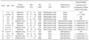
The subjects were seven boys and six girls, aged from 6 months to 7 years 6 months (mean age: 3 years 2 months). There was no significant difference of age (p = 0.93) among the three CT types of tumors. The patients presented with abdominal distension (n = 8), a palpable mass (n = 3) or an incidentally detected tumor during US (n = 2) for other clinical indications: a patient (case 8) with Peutz-Jeghers syndrome, and a patient (case 13) that had a Kasai operation for biliary atresia at one month of age, respectively. The serum level of alpha-fetoprotein, total and direct bilirubin level, alkaline phosphatase level, and level of transaminases (aspartate transaminase and alanine transaminase) were normal in all except two patients that had an elevated level of alpha-fetoprotein: the patient with biliary atresia (case 13) and a 9-month-old girl (case 5). Hepatitis B surface antigen was negative in all patients. In all patients, the tumors were surgically resected. Clinical features of the patients are summarized in Table 1.
All patients had a solitary hepatic tumor. The tumor occurred in the right lobe of the liver in nine patients and in the left lobe in four patients. Splenomegaly, ascites and retroperitoneal lymphadenopathy were noted in one patient that had biliary atresia.
Imaging Characteristics
The longest diameter of the tumors ranged from 1.8 to 20 cm (mean 13.0 cm) and was larger than 10 cm in 11 patients (85%). There was no significant difference in the size of the tumors (p = 0.79) among the three CT types of tumors. The tumor was multiseptated cystic in four patients (31%) (Fig. 1), mixed solid and cystic in five patients (38%) (Figs. 2, 3), and solid in four patients (31%) (Figs. 4, 5).
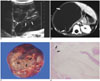
In four patients with a multiseptated cystic tumor, US (n = 1) showed a well-defined tumor with variable cystic spaces and multiple thin septa in the liver (Fig. 1A). All of these tumors showed fine enhancing septa within the cystic tumor on post-contrast CT (Fig. 1B). Neither calcification nor a solid portion was detected in these patients.
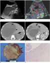
In five patients with a mixed cystic and solid tumor, US (n = 5) showed a well-defined tumor with variable-sized cystic spaces, irregularly thick septa, and a solid portion of variable extent with heterogeneous hyperechogenicity (Figs. 2A, 3A). In three patients, internal debris with fluid-fluid level was seen in the cystic portion of the tumor (Figs. 2A, 2C). On color and power Doppler US (n = 4), linear vascular flow was detected along the thick septa (Fig. 2B) and in the solid portion. On CT, thick septa within the cystic portion (Fig. 2D) and the solid portion of the tumor (Fig. 3C) showed heterogeneous enhancement. A tiny nodular intratumoral calcification was seen in one patient (case 8) (Fig. 3B).
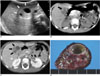
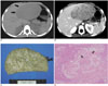
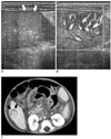
In four patients with a solid tumor, US (n = 3) showed a well-defined, isoechoic tumor (n = 1) or slightly hyperechoic (n =2 ) (Fig. 5A) tumors. Color or power Doppler US (n = 3) showed central (Fig. 5B) or peripheral vascularity in two patients and one patient, respectively. These tumors showed low attenuation on a pre-contrast CT scan in all patients, except for one with a high attenuating lesion. At post-contrast CT, tumors were heterogeneously enhanced in all patients (Figs. 4B, 5C). In the patient that had biliary atresia (case 13), the tumor appeared as a 1.8-cm sized enhancing nodule with a peripheral rim of low attenuation at post-contrast CT (Fig. 5C).
No demonstrable tumor capsule was seen in any patients.
Pathological Correlation
The mesenchymal hamartomas showed multiple variable sized cystic portions within the tumor in their gross morphology (Figs. 1C, 2E, 3D). Each mesenchymal hamartoma with a multiseptated cystic appearance contained various amounts of myxoid or collagenous stroma and showed proliferation of the malformed bile ducts with periductal collaring of the stromal cells (Fig. 1D). All of the four patients with a multiseptated cystic tumor contained a small amount of hepatocytes (< 10%) on a semiquantitative evaluation.
The mesenchymal hamartomas with a mixed solid and cystic appearance also contained various amounts of myxoid or collagenous stroma and showed proliferation of the malformed bile ducts with periductal collaring of the stromal cells (Fig. 2F). Among five patients with a mixed type mesenchymal hamartoma, the amount of hepatocytes was small in three patients and moderate in two patients.
In four patients with a solid mesenchymal hamartoma, a moderate (n = 3) amount or abundant amount (n = 1) of hepatocytes was found in the cords, islands or lobular pattern. The solid tumor also contained various amounts of mesenchymal stroma and proliferation of the small bile duct was seen (Fig. 4D). In one patient with an enhancing nodule with a peripheral rim of low attenuation (case 13), the enhancing portion revealed a large amount hepatocytes histologically with a rim of myxoid stroma. The amount of hepatocytes was significantly different among the multiseptated cystic, mixed solid and cystic, and solid tumors (p = 0.042). The other pathological findings such as the amount of mesenchymal stroma, the presence of a bile duct or vessel proliferation were not statistically significant among the three groups of the different imaging spectrum.
DISCUSSION
A mesenchymal hamartoma is the second most common benign hepatic tumor in children and accounts for 8% of all hepatic tumors in children (10-14). Histologically, a mesenchymal hamartoma appears as a disordered arrangement of mesenchyme, bile ducts and hepatocytes (1-3, 9, 15). Cords of normal-appearing hepatocytes are separated by zones of loose poorly cellular mesenchyme. The porous nature of a mesenchymal hamartoma permits accumulation of fluid. Cystic degeneration of the mesenchyme with resultant fluid accumulation from obstruction and dilatation of the lymphatics and/or by entrapped bile ducts may lead to enlargement of the tumor (12). The margin between the liver and the tumor is distinct; however, a true capsule is generally not present (2). The level of serum alpha-fetoprotein, which is believed to be secreted by the proliferating hepatocytes within the tumor, can be elevated (13, 14).
A multiseptated cystic appearance, which may be a typical finding of mesenchymal hamartoma, is rarely seen in other hepatic tumors in children. Intratumoral calcification, which can be frequently detected in hepatoblastomas (over 50%) or infantile hemangioendotheliomas (up to 40%) (11), has been reported very rarely for a mesenchymal hamartoma. Chung et al. (8) described the CT findings of a mesenchymal hamartoma containing central and peripheral calcifications in a 57-year-old woman. Konez et al. (15) also described the CT findings of the multiseptated cystic form of a mesenchymal hamartoma with a septal calcification in a 10-year-old boy. In this series, a tiny calcification within the tumor was seen only in one patient.
Although a mesenchymal hamartoma present most frequently as a multiseptated cystic or mixed solid and cystic tumor (1-5), it can rarely occur as a solid tumor (6-9). In this study, according to the extent of the cystic component on CT scans, we classified each tumor into one of three types for evaluating the imaging spectrum of this tumor. In this study, the tumor was totally solid in 31% of the patients, and one case of a solid mesenchymal hamartoma (case 11) showed features of a so-called "mixed hamartoma." The mixed hamartoma is a rare hamartomatous lesion composed of all cellular components of the normal liver, including a larger amount of hepatocytes, ductal proliferation, and hepatocellular-ductal transition. The mixed hamartoma and mesenchymal hamartoma may be in one disease spectrum, since there is some overlapping in the histological features of both hamartomas (16, 17). In children with a solid mesenchymal hamartoma, radiological differential diagnosis should include various hepatic tumors such as a hepatoblastoma, a hepatocellular carcinoma, an infantile hemangioendothelioma, a hepatic adenoma, or a focal nodular hyperplasia. Heterogeneous enhancement and absence of a tumor capsule in a mesenchymal hamartoma can be helpful in the differential diagnosis from other tumors.
As in one of our patients that had biliary atresia and a solid mesenchymal hamartoma (case 13), Lack (18) described a mesenchymal hamartoma in a 5-month-old girl with biliary atresia, which was found incidentally during an exploratory laparotomy for obstructive jaundice. The cystic component of this tumor was either very small or undetectable. Lack (18) showed an interest in this case because one of the theories for development of a mesenchymal hamartoma is biliary obstruction (19).
In conclusion, a mesenchymal hamartoma of the liver in children can show a wide spectrum of radiological features from a multiseptated cystic tumor to a mixed solid and cystic tumor, and even a solid tumor.
 XML Download