

 PDF
PDF ePub
ePub Citation
 Print
Print


Stenosis and occlusion of the carotid and cerebral arteries are known to decrease cerebral blood flow and cerebral blood flow reserve. Medical treatment is difficult in such cases, and superficial temporal artery to middle cerebral artery (STA-MCA) bypass surgery has been performed in some centers to treat brain ischemia and prevent new ischemic episodes (1-6). However, the number of STA-MCA surgeries decreased after a consensus study published in 1985 reported that STA-MCA bypass surgery does not significantly reduce the rates of reinfarction (7).
However, a novel method of evaluating cerebrovascular expansion using the vasodilator acetazolamide was introduced after the 1985 consensus report was published. Patients with arterial stenosis or occlusion show little or no improvement in cerebral blood flow in response to acetazolamide. Patients who have abnormal responses to acetazolamide infusion are known to carry a high clinical risk of recurrent cerebral ischemia or infarction after arterial occlusion, which is believed to be due to an insufficient supply of physiologic collateral vessels. Therefore, it is very important to evaluate the vascular reserve in patients with cerebral ischemia (8-10).
Vascular reserve can be assessed by comparing brain perfusion single photon emission computed tomography (SPECT) images before and after the infusion of acetazolamide. Vascular reserve is preserved and blood flow generally increases in response to acetazolamide in most cases where the perfusion pressure is normal and sufficient collateral vessels are established. However, when the collateral vessels are inadequate, the vascular reserve decreases and blood flow does not increase or may even decrease upon the administration of acetazolamide.
The evaluation of baseline and acetazolamide brain perfusion SPECT images relies on visual and semi-quantitative analyses. The inherent subjectivity and inter-reader variability associated with the evaluation of these images make quantitative analyses difficult. Fortunately, imaging technology and image analysis programs have improved in recent years, and a more objective quantitative study has been made possible by the development of statistical parametric mapping (SPM). Using SPM to analyze the resulting images, we attempted to attain a more objective assessment of the effectiveness of STA-MCA bypass surgery by measuring the changes in postoperative cerebral blood flow.
MATERIALS AND METHODS
Subjects and Image Acquisition
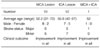
The subjects were patients who were diagnosed with occlusion or at least 80% stenosis of the left internal carotid artery (ICA) and/or left MCA during visits to our neurosurgery department between June 1999 and June 2003 and subsequently underwent left side STA-MCA bypass surgery (Table 1). Only patients with left side occlusion were included in order to ensure an impartial analysis. Patients who had previous operations or a history of head trauma were excluded, as were cases in which acute postoperative complications, such as hemorrhage, occurred.
A total of 23 patients (16 male and 7 female) were included in the study. The patients ranged in age from 31 to 72 years, with mean age of 54.3 ± 10.9 years. Preoperative baseline and acetazolamide brain perfusion SPECT images were obtained from all patients two to three weeks before surgery, with a less than three-day interval between the two SPECT scans. Postoperative baseline and acetazolamide SPECT scans were performed within three weeks of the surgery, and the interval between the postoperative baseline and acetazolamide tests was also less than three days.
All baseline brain perfusion SPECT scans were performed with the patient lying supine on a bed in a dark, quiet room. Forty minutes after 740 MBq of Tc-99m-ECD was infused intravenously, images were taken using a dual-head gamma camera (E-CAM, Siemens, Germany) with a low-energy fan-beam collimator. Acetazolamide brain perfusion SPECT scans were performed under the same conditions by administering 1 gm of acetazolamide intravenously 15 minutes before infusing 1,110 MBq of Tc-99m ECD. Both baseline and acetazolamide-infused images were taken by rotating the camera a total of 360 degrees at 3-degree intervals. Images were taken at a rate of 20 seconds per frame. The data was acquired on a 128 × 128 matrix with a 20% symmetric window at 140 keV. Continuous trans-axial tomograms of the brain were reconstructed after filtered back-projection with a Butterworth filter (cutoff frequency 0.4 cycles/pixel, order 5) to reduce statistical noise. Tc-99m ECD images were corrected for tissue attenuation using a standard, commercial correction routine, which assumes uniform attenuation (attenuation coefficient = 0.15 cm-1).
Image Analysis
Statistical parametric brain maps constructed from images of baseline and acetazolamide-infused brain perfusion SPECTs were analyzed in SPM99 (Statistical Parametric Mapping 99, University College of London, UK) using Matlab (Mathworks Inc., USA). SPECT data with attenuation and scatter correction were converted into Analyze format (Mayo Foundation, Baltimore, MD) with voxel dimensions of 3.9 mm in each spatial dimension. The mean pixel intensity across all slices of the imaging volume was calculated. The pixel threshold was set to 80% of this value to eliminate the background noise and partial volume effects at the edge of the brain. The count of each voxel was normalized versus the total count for the brain (proportional scaling in SPM with a global mean of 50). Each SPECT scan was then spatially normalized by 12-parameter affine warping and tri-linear interpolation to the SPECT template brain from the Montreal Neurological Institute, reformatted to a 16-bit image having a size of 79 × 95 × 68 voxels and a 2 × 2 × 2 mm voxel dimension. These images were smoothed with a Gaussian filter of 16 mm full-width at half maximum (FWHM). Using SPM, total of four paired t-tests were performed in order to compare the following pairs: preoperative baseline and acetazolamide SPECT images, postoperative baseline and acetazolamide SPECT images, preoperative and postoperative baseline SPECT images, and preoperative and postoperative acetazolamide SPECT images. In the SPM analysis, an uncorrected p-value of 0.001 was considered significant, and areas showing more than 100 activated voxels were analyzed. To determine the exact anatomical location of the resulting region overlapping the area on the standardized brain map, the X, Y, and Z coordinates of each voxel were entered into Talairach Daemon software.
RESULTS
Clinical Progress
All patients were followed up for six months to two years after surgery, and none of them experienced recurrence or exacerbation of symptoms. The pre- and postoperative neurological status of each patient was assessed according to the National Institute of Health Stroke Scale (NIHSS). All patients showed improved neurological function after the surgery: 11 patients who showed minor neurological deficits preoperatively experienced improved symptoms after surgery (neurological improvement of > 2 on NIHSS), and 12 patients with major neurological deficits before surgery also showed improvement postoperatively (neurological improvement of > 3 on NIHSS).
Results of Image Analysis
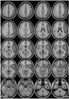
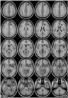
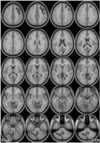
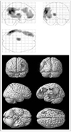
The overall regional cerebral blood flow (rCBF) in the left cerebral hemisphere was decreased on preoperative baseline SPECT images in comparison to that observed on the preoperative acetazolamide SPECT images (Fig. 1). In addition, when the pre- and postoperative acetazolamide SPECT images were compared, the rCBF in the left temporal, frontal, and left cerebellar areas was found to be increased postoperatively (Fig. 2). Similarly, comparison of the pre- and postoperative baseline SPECT images revealed that the rCBF in the left frontal and right cerebellar areas was increased postoperatively (Fig. 3). Finally, the rCBF in the left temporal, parietal, and frontal cortices as well as the thalamus was increased on the postoperative acetazolamide SPECT images in comparison to the baseline SPECT images (Table 2, Fig. 4). On the other hand, on both the pre- and postoperative images, there was no brain area in which the acetazolamide SPECT images showed diminished blood flow when compared to the baseline SPECT images. These findings indicate that mild improvement in the rCBF and response to acetazolamide can be expected after STA-MCA bypass surgery.
DISCUSSION
Some reports have suggested that patients suffering from transient ischemic attacks of the brain or hemodynamically compromised cerebral ischemia may benefit from bypass surgery (4, 11, 12). Considering the operative risks and complications associated with bypass surgery, it is very important to select the patients who are most likely to benefit from bypass surgery.
Several SPECT methods that rely on different radioisotopes, such as baseline and acetazolamide-infused Xe-133, I-123 IMP, and Tc-99m heaxmethyl propylene amine oxime (HMPAO)/ECD brain perfusion SPECT, have been developed in order to assess brain blood flow and reserve. All of these SPECT methods can provide a more accurate measurement of blood flow than conventional angiography. Schmiedek assessed the brain blood flow reserve by measuring brain blood flow at rest and after acetazolamide infusion in patients with occlusive cerebrovascular diseases using Xe-133 SPECT and reported that the blood flow reserve significantly decreased in patients with insufficient collateral circulation. In addition, he stated that while there was little difference in the baseline blood flow reserve before and after external carotid artery-internal carotid artery bypass surgery, the blood flow improved after acetazolamide infusion (4). Kohno et al. used Xe-133 SPECT to evaluate the blood flow and reserve before and after surgery in patients with Moyamoya disease and reported that the blood flow reserve in these patients significantly improved after surgery (13). These results prove the efficacy of surgery in hemodynamically compromised patients with arterial stenosis. This study using Xe-133 SPECT analysis also illustrated the efficacy of surgery more objectively than CT or angiography. Though actual clinical symptoms should be the golden standard, clinical judgments can be very subjective.
Blood flow reserve assessed by brain perfusion SPECT is associated with the oxygen extraction rates measured by PET. Powers et al. studied the consequences of bypass surgery in patients with chronically depressed local brain perfusion pressure using PET and found this imaging modality to be ineffective (14). Schmiedek stated that the degree of hemodynamic compromise was inconsistent among the subjects and that PET, which primarily measures the capacity for auto-regulatory vasodilation, is not an appropriate modality for measuring the success of the bypass surgery (4). Therefore, SPECT is more effective than PET for evaluating brain perfusion and blood flow reserve.
Being able to quantify the degree of improvement in perfusion, whether between different groups or in an individual, is very important in nuclear medicine scintigraphy. However, it is extremely difficult to compare the results of quantification, whether in a single subject undergoing serial studies or in multiple subjects pooled into groups. Qualitative analysis is usually limited to the area of interest, but such studies invariably suffer from low accuracy and poor reproducibility. A number of methods have been developed in order to overcome these limitations, and SPM is one such method that is commonly used.
Lee et al. performed baseline and acetazolamide-infused Tc-99m HMPAO SPECT in patients undergoing STA-MCA surgery and analyzed the pre- and postoperative results using SPM and SPAM (statistical probabilistic anatomic map) (15). They reported that a general improvement of baseline brain blood flow was distributed over the left hemisphere of the cerebrum, including the frontal, temporal, parietal, and occipital lobes, where the operation was performed. The reserve capacity increased in the superior temporal and posterior parietal areas. In addition, comparison with a control group demonstrated that the blood flow in the superior temporal and posterior parietal areas increased after surgery.
We used Tc-99m ECD to evaluate brain perfusion, and baseline and acetazolamide SPECT images were obtained on different days within a lag period of three days. In addition, in contrast to other studies, baseline and acetazolamide SPECT scans were performed both pre- and postoperatively in the patient group. All paired sets of data were compared and displayed as parametric images. Comparison of the baseline and postoperative acetazolamide images showed increased blood flow at the operation site, which included the temporal, frontal, and left cerebellar areas, which is believed to have resulted from the effects of surgery and changes in vessel reserve following acetazolamide infusion. In particular, the acetazolamide SPECT images illustrated the general increase in the blood flow in the left cerebrum very well. In addition, the baseline blood flow after STA-MCA anastomosis appeared to increase in the right cerebellum based on comparison with the preoperative baseline studies, and this increased blood flow seems to be the result of improved crossed cerebellar diaschisis after bypass surgery. Furthermore, the relationship between the baseline and acetazolamide infusion images following surgery revealed that the circulation in the anastomosis site and small vessels around the anastomosis site was not greatly affected by the degree of decrease in resting state brain blood flow or the level of response to acetazolamide infusion, which is in agreement with result reported by Iwama et al. (5). Collateral circulation through the anastomosis site is probably more dependent on the degree of arterial stenosis and surgical technique. In summary, this study demonstrates that by forming adequate collateral circulation around the anastomosis site after bypass surgery in patients with ischemia due to aberrant hemodynamics, a significant increase in response to acetazolamide, or blood flow reserve, may be achieved even when the baseline flow remains unchanged. This finding agrees with those of many previous reports and was confirmed more objectively in this study than in the previous studies.
This study has a number of limitations. One limitation of the present study is that a correlation analysis with more detailed clinical data was not performed to evaluate the direct effects of the bypass surgeries. This study is also limited by the fact that individual patient analysis and quantitative evaluation were not performed. Further research with additional clinical data and individual analyses are required in order to confirm the findings of this study.

 XML Download