

 PDF
PDF ePub
ePub Citation
 Print
Print


Aspergillosis is a rare cause of spondylitis, and early diagnosis by MR imaging and adequate treatment are essential for a good outcome (1-3). Although the MR findings of bacterial spondylitis have been fully described (4, 5), the findings of aspergillus spondylitis have been rarely described, and to the best of our knowledge multilevel involvement of cervico-thoraco-lumbar spine has not been previously reported. Here, we report the MR imaging findings of aspergillus spondylitis involving the cervico-thoraco-lumbar spine in a liver transplant recipient.
CASE REPORTS
A 46-year-old man underwent liver transplantation due to hepatitis B virus cirrhosis in March 2005, and subsequently was treated using routine immunosuppression therapy. However, his early postoperative course was complicated by pulmonary aspergillosis. About 10 weeks after liver transplantation a left lower lobe wedge resection and pathology showed aspergilloma, and about three weeks after this thoracic surgery the patient complained of back pain. Plain radiography of the lumbar spine demonstrated no abnormality, and although his back pain was treated conservatively, the patient complained of progressive back pain.
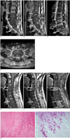
MR imaging of the lumbar spine revealed band-like or diffuse hypointense signals in vertebral bodies L2 to L5 on T1-weighted images (Fig. 1A), which were isointense to slightly hyperintense on T2-weighted images (Fig. 1B). In detail, T1-weighted images showed some hypointense signals with preservation of intranuclear clefts in the L2-3 and L4-5 discs, but disc hyperintensity was absent and intranuclear cleft loss was visualized in the L3-4 disc. In addition, endplate irregularities were apparent in the involved spine. Disc space narrowing was observed at L3-4 and L4-5, and band-like or diffuse enhancement was observed in involved vertebral bodies with an epidural abscess (Fig. 1C). A paraspinal abnormal signal was relatively well-defined (Fig. 1D). The MR based diagnosis was of tuberculous spondylitis rather than pyogenic spondylitis. At surgery, infected granulation tissue and disc material with pus were found at the L3-4 and L4-5 levels, and osteolysis was observed at the inferior endplate of L3, the superior and inferior endplates of L4, and the superior endplate of L5. After curettage and debridement of these lesions, an iliac crest bone block was inserted. Histologically, acute inflammation and necrosis with fungal hyphae suggested aspergillus infection. Histopathologically periodic acid-Schiff and Gomori methenamine silver staining revealed aspergillus infection.
About three months after he underwent lumbar spine MR imaging, he was readmitted for posterior neck pain. MR images of the cervical and thoracic spine showed changes in bone marrow and endplates at the C4-5 and T2-4 levels, the latter of which were similar to the previous findings of lumbar involvement (Figs. 1E-G). Increased disc signal and loss of intranuclear cleft were observed in C4 5 and T2-4 discs. Endplate irregularities and disc space narrowing were observed in the involved spine. During surgery on the cervical spine, infected granulation tissue and disc material were found at the C4-5 level with osteolysis at the inferior endplate of C4 and the superior end plate of C5. Debridement and bone grafting were performed, and pathologic reports disclosed acute inflammation with necrosis and osteomyelitis. Histopathological periodic acid-Schiff and Gomori methenamine silver staining findings revealed aspergillus infection (Figs. 1H, I).
DISCUSSION
Invasive aspergillosis is a life threatening fungal infection that is associated with a high mortality rate despite treatment. Symptoms and signs are nonspecific. In previous reports times between symptom onset and a definite diagnosis were of the order of months (4-7), and in our patient, symptoms persisted for more than two months before pathologic diagnosis. Ordinary laboratory findings are of little help in the diagnosis of aspergillosis (6).
According to a previous report by Williams et al. (8), the absence of disc hyperintensity and intranuclear cleft preservation on T2-weighted images are suggestive of nonpyogenic spondylitis. In our case, decreased disc signal intensity was observed in three of six affected discs and intranuclear clefts were preserved in two of six affected discs. The minimal hyperintensity or isointensity of vertebrae on T2-weighted images observed in our case is consistent with the findings of previous reports (8, 9) on Candida or Aspergillus spondylitis.
Aspergillus spondylitis is often confused with tuberculous spondylitis (3, 10). In a previous case report (3), MR showed involvements of three consecutive vertebral bodies and a well-defined paraspinal mass, and as a result, tuberculous spondylitis was initially considered. Finally, the patient was pathologically confirmed as having Aspergillus spondylitis. Based on our experiences, the following MR findings favor tuberculous spondylitis rather than pyogenic spondylitis; a well-defined paraspinal abnormal signal rather than an ill-defined paraspinal abnormal signal, a thin and smooth abscess wall rather than a thick and irregular abscess wall, the presence of a paraspinal or an intraosseous abscess, subligamentous spread over at least three vertebral levels, multiple vertebral body involvement, and thoracic spine involvement (5). In the present case, four consecutive vertebral bodies were involved and an abnormal paraspinal signal was relatively well-defined on the initial lumbar spine MRI, which indicated tuberculous spondylitis rather than pyogenic spondylitis. However, no paraspinal or intraosseous abscess was evident despite the extensive involvements of four consecutive vertebral bodies and an abnormal paraspinal signal. Because based on our experiences, about 95% (15/20) of tuberculous spondylitis cases have a concomitant paraspinal or intraosseous abscess (5), the absence of a paraspinal or intraosseous abscess appears to be unusual in tuberculous spondylitis. However, as South Korea is still an endemic area for tuberculosis, and initial diagnosis of tuberculous spondylitis was made despite some inconsistent MR findings. In retrospect, we should have considered a fungal infection, such as, Candida or aspergillus spondylitis in this immunocompromised patient.
In conclusion, aspergillus spondylitis should be considered in the differential diagnosis of immunocompromised patients with MR findings resembling those of tuberculous spondylitis.
 XML Download