

 PDF
PDF ePub
ePub Citation
 Print
Print


Although the epidermal inclusion cyst is an uncommon finding in the breast, we can easily diagnosis this as a cyst. But when it is presented in an unusual subareolar location and with a ruptured state, it can be mistaken for breast malignancy. We present here two surgically confirmed cases of ruptured epidermal inclusion cyst in a subareolar location, and this has not been previously described in the English medical literature.
CASE REPORTS
Case 1
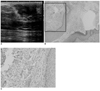
A 50-year-old woman presented with left subareolar pain and a bean-sized palpable mass for one week. Sonography reveals an ill defined heterogeneous, hypoechoic, irregular shaped 1.8×1.1-cm mass in the left subareolar region (Fig. 1A). Fine needle aspiration biopsy (FNAB) showed copious eosinophils, small amounts of ductal epithelium and no malignant cells. According to the patient request, excision was performed and the mass was pathologically confirmed as a ruptured epidermal inclusion cyst (Figs. 1B, C).
Case 2
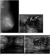
A 44-year-old woman presented with left periareolar pain for several months. Mammography revealed left subareolar asymmetry, periareolar skin thickening and axillary lymph node hypertrophy (Fig. 2A). Sonography shows a 2.2×1.7-cm ill defined mass with an irregular shape, heterogeneous echogenicity and posterior enhancement (Fig. 2B). FNAB was performed to differentiate this from inflammatory breast cancer, but malignant cells were not found. The symptoms were improved after administering antibiotics, and we formed a clinical impression of breast abscess. One year later, the patient presented again with yellowish discharge of the left nipple. On the follow up mammogram, the density of the left subareolar asymmetry and skin thickening was decreased, but it was still observed, and no significant change was observed in the left axillary lymph nodes. On sonography, the previously observed mass showed decreased size (1.5×1.5-cm) and echogenicity. One month after antibiotic administration, the mass disappeared and an irregularly shaped hypoechogenicity was seen on the follow-up sonogram (Fig. 2C). About seven months later, the patient presented with a heat sensation and pain on the same area. The previous hypoechogenicity area changed to a 1.1×0.8-cm mass on sonography and a recurrent abscess was suspected (Fig. 2D). The mass was excised and pathologically confirmed as a ruptured epidermal inclusion cyst.
DISCUSSION
Less than 10% of epidermal inclusion cysts occur in the extremity and an even lower number occur in the palms, sole, and breast. Most breast epidermal inclusion cysts occur in the skin layer, but there is a report of occurrence in the breast parenchyma (1).
Epidermal inclusion cysts can be congenital, or they can occur after trauma, reduction mammoplasty (2) and breast augmentation (3). There is also a possibility of metaplastic lesions from columnar cells that have transformed into squamous cells, and there has been one reported case where the hair follicles or pores are obstructed and inflammatory downward growth of the epidermis made an inclusion cyst like the ones formed from sebaceous glands. Gerlock reported two cases of breast epidermal inclusion cysts associated with FNAB (4). Diverse complications can occur with epidermal inclusion cysts, like spontaneous rupture and the development of squamous cell cancer (5). In spontaneous rupture, these cysts release nonabsorbable keratin that acts as an irritant leading to secondary foreign body-type reactions, granulomatous reactions or abscess formation. Some authors have reported Paget's disease arising from not only the nipple epidermis, but also from perinipple epidermal inclusion cysts (6). Asymptomatic lesions do not require treatment, and biopsy is unnecessary if typical sonographic and physical examination findings are found. However, in cases presenting with palpable breast lesions, the patients are often concern about lumps and may request excision. Although a palpable breast mass shows benign findings on mammography, if the sonographic findings need to be differentiated from a well defined breast malignancy, then biopsy is necessary. To prevent inflammatory and malignant change, surgical intervention may be appropriate if circumstances require it. Several different entity diseases or periductal mastitis, abscess, retroareolar malignancies, intraductal papillary lesion and rarely Paget's disease could have caused the symptoms of our cases on initial presentation. But after sonography, the differential diagnosis of abscess and malignancy were taken into accounts (7, 8).
In our cases, we first considered the possibility of breast malignancy because the masses presented as an irregular mass on the initial sonography, and the patients were over the age 40 and we didn't take the possibility of abscess from ruptured epidermal inclusion cyst into consideration due to its rare occurrence and the unusual lesion location. FNAB and follow up imaging study after medical treatment, or the recurrent feature were the ways to later narrow the differential diagnosis. In conclusion, when a subareolar lesion has findings on sonography that are suspicious of malignancy, the differential diagnosis should include a ruptured epidermal inclusion cyst, with or without evidence of inflammation.
 XML Download