

 PDF
PDF ePub
ePub Citation
 Print
Print


Hemorrhagic fever with renal syndrome (HFRS) is an acute infectious disease caused by hantavirus. HFRS is clinically characterized by fever, renal failure and hemorrhage in organs such as lung, kidney, spleen and the pituitary gland (1, 2). Renal medullary hemorrhage is a well-known complication in the kidney (3), but spontaneous rupture of the kidney and perirenal hematoma in HFRS is rare (4, 5), and patients showing continuous bleeding and massive perirenal hematoma have often been surgically treated (6). We report here on a case of HFRS complicated by massive perirenal hematoma, and the patient was treated with transcatheter arterial embolization.
CASE REPORT
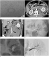
A 48-year-old man visited emergency department due to left lower abdominal pain and oliguria he had suffered with for three days and two days, respectively. He had experienced fever, chills and headache for one week prior to admission. The physical examination on admission revealed petechial rashes on the whole body (Fig. 1A) and tenderness in the left lower abdomen and left flank. He wasn't taking any antiplatelet drugs. The laboratory examination revealed leukocytosis, mild anemia, thrombocytopenia, increased serum creatinine and hyperkalemia. The prothrombin time and partial thromboplastin time were normal.
Contrast-enhanced CT scan of the abdomen and pelvis showed extravasation of the contrast media and massive perirenal hematoma that extended into the pelvic cavity (Figs. 1B, C). Renal arteriography was performed because of the active bleeding and massive perirenal hematoma on the CT scan. Selective left renal arteriography (Fig. 1D) and superselective renal arteriography with a 2-Fr microcatheter (Progreat, Terumo, Tokyo, Japan) (Fig. 1E) showed extravasation of the contrast media from small branches of the renal artery at the inferior portion of the kidney. There was no hypervascular mass or arteriovenous malformation. Transcatheter embolization with Tornado microcoils (Cook, Bloomington, IN) was performed at the ruptured small branches. Renal arteriography following embolization confirmed there was no more extravasation of the contrast media (Fig. 1F). The hemoglobin was 8.9 g/dL on the second day of hospitalization and two pints of packed RBC were transfused. On the third day of hospitalization, the antibody for Hantavirus was found to be positive (the antibody titer to Hantavirus was 1:8192), and the patient was confirmed as having HFRS complicated by perirenal hematoma. Hemodialysis was performed from the first day to the 12th day of hospitalization because of combined renal failure. Follow-up CT scan on the 12th day of hospitalization showed a decreased amount of perirenal hematoma. After recovery, the patient was discharged from the hospital on the 25th day of hospitalization.
DISCUSSION
Hemorrhagic fever with renal syndrome is a rodent-transmitted disease caused by Hantavirus. Rodents can show Hantavirus infection as an asymptomatic carrier state. Transmission to humans occurs by direct contact or inhalation of rodent excrement. Many patients with HFRS show fever, renal failure and a hemorrhagic tendency. The most common manifestation of hemorrhage is petechia involving the skin and oropharyngeal mucosa. Hemorrhage occasionally occurs spontaneously or via minor trauma in such vital organs as lung, kidney, spleen, brain and the pituitary gland (1, 2, 5).
The primary pathologic findings of the kidney in HFRS are renal swelling, intense congestion and hemorrhage in the medulla, and also acute tubular necrosis (3). However, spontaneous renal rupture and perirenal hematoma are rare complications of HFRS. Cho et al. (4) reported two patients with spontaneous perirenal hematoma among 417 patients with HFRS. Guang et al. (5) reported that spontaneous retroperitoneal hematoma occurred in approximately 0.6% of 309 patients with HFRS. Unlike the previous cases, active extravasation of contrast media was revealed on selective renal arteriography in our case.
Vascular endothelial cells appear to be the primary site of Hantavirus multiplication. Virus-induced vascular injury, thrombocytopenia and disseminated intravascular coagulation may cause diffuse hemorrhage, including spontaneous perirenal hematoma (1, 6).
Patients with HFRS who are complicated by active bleeding and massive perirenal hematoma have often been treated with operation. Valiakhmetov et al. (7) reported on 29 patients with HFRS complicated by spontaneous rupture of the kidney and retroperitoneal hematoma. Among them, 11 patients were treated with conservative therapy and 18 patients were treated with operation. The indications for surgery were progressive anemia, palpable retroperitoneal hematoma and symptomatic peritoneal irritation. All patients underwent evacuation of blood clots and ligation of the ruptured branches of the renal artery. However, in our case, we performed superselective renal artery embolization instead of operation. To the best of our knowledge, there has been no reported case treated with transcatheter arterial embolization.
Spontaneous renal bleeding and perirenal hematoma have various causes. The well-known causes are renal cell carcinoma, angiomyolipoma, vascular causes such as renal infarction and arteriovenous malformation (AVM), and other causes (hemorrhagic cyst, hemorrhagic abscess and idiopathic hemorrhage) (8, 9). HFRS could be included in the differential diagnosis if there is no mass or vascular abnormality on the imaging studies of a patient with acute spontaneous perirenal hematoma. The MR imaging of the kidneys may be helpful for the differential diagnosis because low signal intensity in the outer medulla on the T2 weighted images is a characteristic finding of medullary hemorrhage in HFRS (3).
In summary, spontaneous rupture of the kidney and perirenal hematoma is a rare complication of HFRS. We report here on a case of HFRS that caused massive perirenal hematoma, and this was treated with superselective renal artery embolization.
 XML Download