

 PDF
PDF ePub
ePub Citation
 Print
Print


Primary tumors of the seminal vesicles are rare. Primary adenocarcinoma of the seminal vesicles commonly occurs in patients who over the age of 50. Congenital anomalies of the mesonephric duct are rare in males. Congenital seminal vesicle cysts are commonly associated with ipsilateral renal agenesis or dysgenesis (1). We present here the imaging findings of a primary adenocarcinoma of a seminal vesicle cyst associated with an ectopic ureter opening into a seminal vesicle and ipsilateral renal agenesis.
CASE REPORT
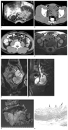
A 41-year-old man presented with terminal gross hematuria that was noted one month earlier. His past medical history was unremarkable except that he had suffered with prostatitis. The serum markers for prostate cancer, prostate-specific antigen (PSA) and prostatic acid phosphatase (PAP) were normal. Carcinoembryonic antigen (CEA) was also in the normal range. Transrectal ultrasonography showed a well-demarcated, pear-shaped cystic lesion that included an echogenic papillary solid mass in his left seminal vesicle (Fig. 1A). Contrast-enhanced pelvic CT showed that the papillary solid mass originated from the wall of the left seminal vesicle cyst and it was mildly enhanced (Fig. 1B). A dilated ectopic ureter opening into the dilated left seminal vesicle was also seen. There was no evidence of any other pelvic tumors. Contrast enhanced abdominal CT at the level of the L4 vertebra demonstrated a small abnormal soft tissue density in the aorta's left lateral aspect, suggesting a dysgenetic or atrophic kidney (Fig. 1C). The axial T1-weighted (TR/TE: 540/12 ms) MR image showed high signal-intensity fluid in the seminal vesicle cyst (Fig. 1D). The coronal T2-weighted (TR/TE: 5500/136 ms) MR image demonstrated an approximately 7.8 × 6 × 5.2 cm papillary mass in the left seminal vesicle cyst (Fig. 1E). The sagittal T2-weighted image showed a markedly dilated ectopic ureter draining into the cyst (Fig. 1E). The cystoscopic findings showed bulging of the left hemitrigone on the left side of the bladder. The left ureteral orifice was absent.
Radical excision of the left agenetic kidney, left ureter and seminal vesicle cyst and a partial cystectomy were done. The seminal vesicle cyst contained a movable solid mass. The gross specimen showed the internal surface of the seminal vesicle cyst (Fig. 1F). The main papillary mass was removed. Photomicrography showed a papillary glandular configuration covered with carcinoma cells and mucinous materials in the cystic space without any muscular invasion (Fig. 1G). The histopathologic diagnosis was a well-differentiated primary mucinous adenocarcinoma of the left seminal vesicle cyst (Fig. 1H). The specimen we labeled "agenetic kidney" showed only a vestigial remnant of the ureteric buds. Five years later, the follow-up CT and whole body bone scanning revealed no evidence of tumor recurrence or distant metastasis.
DISCUSSION
Primary tumors of the seminal vesicles, such as adenocarcinoma, sarcoma or lymphoma, are rare findings, and secondary tumors are more common. Most often, the patients present late in their disease course with nonspecific symptoms (2), so a late diagnosis of primary adenocarcinoma in the seminal vesicle is rather typical. A mass is often palpable above the prostate and it is usually nontender. Our patient had a previous history of prostatitis seven years ago.
Elevated serum levels of PSA and PAP helped us to identify the prostate as the site of primary malignancy. On the other hand, the serum PSA and PAP levels are normal in patients with primary seminal vesicle adenocarcinoma, but the serum CEA may be elevated (3). In our patient, all of these serum markers were normal.
Ureteral ectopia more commonly develops in females than in males (5). The mesonephric duct extends caudally to the cloaca and it gives raise to the ureteric bud. If the ureteric bud originates more cranially than normal in the mesonephric duct, then the ureter may insert into seminal vesicle, the posterior urethra, the ejaculatory duct and the vas deferens (2). Ureteral ectopia in males usually involves drainage into the seminal vesicle (5). Ectopia in these areas may be associated with reflux or obstruction, and this is the suggested cause of renal functional impairment or absence (4). So, seminal vesicle cysts are commonly associated with renal agenesis or dysgenesis on the ipsilateral side (6). Incomplete development between Wolffian's duct and the urogenital sinus in males results in an accumulation of secretions and the subsequent formation of seminal vesicle cysts during puberty (7). We suggest that the cause of large seminal vesicle cysts is not only the ectopic ureter opening into the cyst, but also a mucin producing tumor.
Secondary involvement of the seminal vesicle is more common than primary seminal vesicle cancer for carcinoma in situ of the bladder, adenocarcinoma of the prostate, lymphoma or rectal carcinoma. The diagnosis of primary adenocarcinoma within the seminal vesicle is difficult to arrive at with using just the image findings. But in our patient, the intraluminal protruding papillary mass in the seminal vesicle cyst was easily identified on imaging study. We suggest that primary seminal vesicle cancer should be considered in the differential diagnosis of papillary mass in the cyst.
Okada et al. have reported a case of papillary adenocarcinoma in a seminal vesicle cyst associated with ipsilateral renal agenesis; however, an ectopic ureter opening was not described (8). A few cases of ectopic ureter associated with cancer have been previously reported (8, 9).
Primary seminal vesicle adenocarcinoma commonly occurs in patients over the age of 50. Most patients with seminal vesicle cysts combined with ipsilateral renal agenesis or dysgenesis are asymptomatic until the condition's second or third decade (1, 2). Our patient was younger than the patients in the previously reported cases who had primary seminal vesicle adenocarcinoma discovered, and he was older than the patients who had seminal vesicle cysts combined with ipsilateral renal agenesis or dysgenesis. So, we suggest the possibility that the development of seminal vesicle adenocarcinoma is due to chronic stimulation of the ectopic ureter's secretion. In this clinical setting with a papillary mass of a seminal vesicle cyst with an ectopic ureter, primary mucinous adenocarcinoma arising from the seminal vesicle cyst should be considered as part of the differential diagnosis, even though the patient was young.
In conclusion, we report here on a case of primary mucinous adenocarcinoma arising from a seminal vesicle cyst that was associated with an ectopic ureter opening and ipsilateral renal agenesis, which is a very rare condition indeed. The lesion was depicted on transrectal ultrasonography, contrast enhanced CT and MRI as a papillary solid mass originating from the wall of the left seminal vesicle cyst.
 XML Download