

 PDF
PDF ePub
ePub Citation
 Print
Print


Varices are most commonly noted in the esophagus and gastric fundus, and they may have ectopic locations such as duodenum, jejunum, ileum, colon, anorectum, retroperitoneum and rarely at the site of colostomy or ileostomy. Ectopic varices are an uncommon cause of gastrointestinal hemorrhage, but they account for up to 5% of all variceal bleedings (1).
Bleeding from stomal varices has been reported in up to 20% of the patients suffering with chronic liver failure with permanent stoma (2). However, the diagnosis of stomal varices is difficult because bleeding from stoma may also be associated with lower gastrointestinal bleeding.
The purpose of this report is to describe the 2D reformatted and 3D volume rendered images of a patient with an episode of acute bleeding from stomal varices. To the best of our knowledge, the 2D reformatted and 3D volume rendered images by MDCT for visualization of ectopic stomal varices have not been previously reported in the medical literature.
CASE REPORT
A 66-year-old man was admitted to our hospital for evaluation of hematochezia into his colostomy bag. He had a history of abdominoperineal resection for rectal cancer that was performed seven years earlier and he also had alcoholic liver cirrhosis (Child class C). On the physical examination, the conjunctivae were anemic, but the abdomen was flat. The pulse was 104 beats per minute and it was regular, the temperature was normal and the blood pressure was 90/50 mmHg. The laboratory values revealed decreased hemoglobin levels (7.2 g/dl), a decreased hematocrit (19.9%) and a decreased platelet count (61,000/µL). The focus of bleeding was thought to be in the superior area of the peristomal skin. Gastroscopy and colonoscopy were performed to exclude that the focus of bleeding was of a intraluminal origin, and the examinations did not reveal any sign of acute bleeding.
The computed tomography images of the abdomen and pelvis were acquired using a 16 detector multislice helical scanner (Sensation 16; Siemens, Germany). The patient was placed in a supine position, and intravenous contrast was infused at a rate of 3 cc/sec, and the imaging performed after a 75 second delay. The study was performed using 120 kV, 140 mA, 0.75 mm beam collimation and a 10 mm per rotation table speed. The images were obtained from the region between just above the level of the diaphragm and immediately below the pelvis.
All the images were stored in the DICOM (Digital Imaging and Communications in Medicine) format. The digital data were sent to a PACS server (π view; Infinitt, Seoul, Korea), and the transverse source images were transferred to a computer workstation that used commercially available software (Rapidia, Infinitt, Seoul, Korea) for 2D multiplanar reformations and 3D volume rendering images.
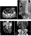
On the 2D reformatted axial and sagittal images, the stoma and the peristomal skin were thickened with the adjacent collateral vessels filled (Figs. 1A, B). The tortuous collateral vessels that drained into the portal vein through the inferior mesenteric vein were well demonstrated on the 3D volume rendering images (thick slab coronal view, Figs. 1C, D). Other collateral vessels were also observed inferiorly, and these vessels drained to the left inferior epigastric vein.
Suture ligation of the tortuous engorged mesocolonic vessels was performed at the superior aspect of the colonic stoma. The bleeding stopped after the procedure, and the patient showed stable vital signs. Although further procedures such as Transjugular Intrahepatic Portosystemic Shunt (TIPS) were needed to manage the recurrent bleeding, the patient and his family refused this procedure. Therefore, the patient was discharged with a warning that rebleeding could occur.
DISCUSSION
Ectopic varices account for between 1% and 5% of all variceal bleedings (1). Typically, they are associated with portal hypertension, and the varices can develop almost anywhere in the gastrointestinal tract. Although ectopic varices can occur at several sites, bleeding from ectopic varices is most commonly found in the duodenum and at sites of previous bowel surgery, including the stoma (3).
Bleeding from stomal varices has been noted in up to 25% of patients with chronic liver disease (liver cirrhosis) with permanent stoma (2). However, the diagnosis of stomal varices is difficult because stomal bleeding cannot be distinguished from gastrointestinal bleeding. Stomal varices are most commonly encountered in patients with history of colostomy or ileostomy for treating inflammatory bowel disease or cancer, and particularly in patients who had proctocolectomy preformed for chronic ulcerative colitis in association with primary sclerosing cholangitis (4). Less often, stomal varices occur in association with ileal conduit (5). Ectopic varices typically develop gradually after the initial operation (6). Stomal varices are characterized by a purplish hue around the stoma.
The management of ectopic stomal variceal bleeding varies widely from simple local measures such as direct pressure dressing, sclerotherpy, epinephrine-soaked gauze, gel foam or suture ligation to surgical interventions such as revision of the stoma, portosystemic shunts or liver transplantation. Moreover, the application of radiological interventions for the treatment of esophageal varices has been extended in recent years to symptomatic stomal varices (3). The use of TIPS to treat ectopic varices was first reported in 1994 for patients with intestinal varices. TIPS is currently considered to be the treatment of choice when dealing with stomal varices in patients with liver cirrhosis and portal hypertension (7). Moreover, because many patients with stomal varices are candidates for liver transplantation, TIPS is preferred to surgical portosystemic shunt.
In our case, the stomal varices were easily detected by the contrast enhanced 2D reformatted and 3D volume rendering images during the portal venous and equilibrium phases of contrast administration. The postprocessing time required for the 2D reformatted and 3D volume rendering images is minimal. Once the axial slices become available at the workstation, a skilled operator can produce the 2D reformatted and 3D volume rendering images in approximately 10 minutes, with commercially available software and using the preset protocols.
Considering the incidence of ectopic stomal varices, the stoma should be included in the CT evaluation of liver cirrhosis patients with permanent stoma. Knowledge of this entity is important when providing care for patients with portal hypertension, because the diagnosis and management of ectopic varices differ from those of esophagogastric varices. Therefore, the 2D reformatted and 3D volume rendering images by MDCT are a useful modality for the evaluation of ectopic stomal varices because MDCT provides good quality images and it is a promising noninvasive imaging tool for the morphologic assessment of abdominal vasculature, including the arterial system as well as the portal vein and its tributaries.
 XML Download