Abstract
Objective
We wanted to describe the characteristic ultrasonography (US) features and clinical findings for making the diagnosis of subacute granulomatous thyroiditis.
Materials and Methods
A total of 31 lesions from 27 patients were confirmed as subacute granulomatous thyroiditis by US-guided fine needle aspiration biopsy. We analyzed the ultrasonographic findings such as the lesion's size, margin and shape, the discrepancy between length and breadth and the vascularity. The clinical findings such as acute neck pain or fever were reviewed. The follow-up clinical and ultrasonographic data were reviewed for 15 patients.
Results
The thyroid gland was found to be enlarged in five patients, it was normal size in 20 patients and it was smaller in two patients. All the lesions had focally ill-defined hypoechogenicity. Hypervascularity was not noted in any of the lesions. Painful neck swelling was present in 18 patients. An accompanying fever was documented in nine of the 18 patients. Twelve patients showed disappearance (n = 3) or a decreased size (n = 9) of their lesions on follow-up US.
Subacute granulomatous thyroiditis is one of the inflammatory thyroid diseases known as de Quervain's disease. This is an uncommon disease that most often occurs in middle-aged women. The disease affects women three to five times more often than men, and viral infection is the most common cause of the disease (1, 2). Subacute granulomatous thyroiditis can be diagnosed clinically, and the features of the disease include acute thyroid pain, hypermetabolism, a low grade fever, occasional dysphagia, suppressed levels of thyroid stimulating hormone (TSH), and an elevated erythrocyte sedimentation rate (3). A painful thyroid following an upper respiratory tract infection is often a sign of subacute granulomatous thyroiditis. This self-limited disease often resolves spontaneously, usually without subsequent thyroid function abnormalities (4), or it may improve with anti-inflammatory medications and corticosteroids, and often within 24 hours (5).
Thyroiditis is generally clinically diagnosed after evaluating both the clinical and laboratory data. This is because the characteristic ultrasonographic features that suggest thyroiditis have not been clearly defined (6). However, several recent reports have shown that ultrasonography (US) is a useful tool for diagnosing and monitoring thyroid pathology, including thyroiditis (7-9).
In some cases, the thyroid gland has been reported to be enlarges to approximately twice its normal size (9). Some of the other ultrasonographic findings are hypoechoic areas and a lack of flow on color Doppler US (1). In this study, we tried to determine the characteristic ultrasonographic and clinical features of subacute granulomatous thyroiditis.
Between January 2000 and May 2003, we reviewed the medical records at our institute and we found 4,672 thyroidal cytological reports that were based on US-guided fine needle aspiration biopsy (FNAB). A total of 31 lesions from 27 patients were found to exhibit subacute granulomatous thyroiditis. Four patients had two lesions that were biopsied at the same time. Most of the aspiration biopsies were recommended by radiologists because of the difficulty in differentiating subacute granulomatous thyroiditis from thyroid malignancy. Some of the biopsies were requested asked by other physicians or by the patients because of concern about the lesions. All the aspiration biopsies were performed with 23 G needles; this was done twice for each lesion at the same session. Twenty-five patients were female and two were male. The patients were between 29 and 70 years of age (mean age: 48.2 years).
Ultrasonography was performed using an HDI 3000/5000 ultrasound scanner (Advanced Technology Laboratories, Bothell, WA) equipped with a 12-MHz linear-array transducer. The US findings were analyzed radiologists working in consensus. The clinical findings such as acute neck pain or fever were reviewed from the medical records. Follow-up evaluations of the ultrasonographic and clinical features were available for 15 patients.
Mild to severe painful neck swelling was present in 18 patients, and an accompanying fever was documented in nine of these 18 patients. Even after reading the medical records, it was not always certain whether or not an upper respiratory tract infection occurred before the onset of the subacute granulomatous thyroiditis. The initial laboratory data at the time of diagnosis showed an elevated ESR (15-55 mm/hr, mean: 37 mm/hr [normal, 0-20.0]) in 12 of the 16 examined patients.
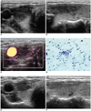
On gray-scale US, unilateral thyroid involvement was demonstrated in 23 patients, and bilateral involvement was demonstrated in four patients. The thyroid gland was enlarged more than 1.8 cm at the anterior-posterior diameter in five patients, it was normal sized in 20 patients and it was decreased in size in two patients. Of the five patients with enlarged glands, four had unilateral enlargement and one had bilateral enlargement. Focally ill-defined heterogeneously hypoechoic areas (the largest diameter: 8-26 mm, mean: 17 mm) were found in all lesions (Figs. 1, 2). Nine of these patients had an irregular or microlobulated margin, and nodular space-occupying hypoechogenicity that mimicked thyroid carcinoma (Fig. 3). The lesions had a tendency to be long (i.e. not round or ovoid shape) on the longitudinal scan, unlike the transverse scan on which they appeared as focally ill-defined masses. The ratio between the length and the breadth of the lesions was 0.8-2.2 (mean: about 1.4). No hypervascularity was noted in any of the lesions on color Doppler US.
Fourteen patients received steroid therapy. Follow-up US was available after therapy for 10 patients. Two patients had no remaining lesions. The lesions were decreased in six patients, and no change in lesion size was seen in two patients. Four of five patients with enlarged glands had a reduced lesion volume. Among the 13 patients who had not been treated, the follow-up data were available for five patients. Of these patients, the lesion had completely resolved in one patient, and the lesions had decreased in size in three patients.
Subacute granulomatous thyroiditis is a self-limited thyroid disorder that presents with localized neck pain and/or low-grade fever. The basic clinical and laboratory data are needed for making the diagnosis, and a biopsy is rarely required (6). The number of diagnoses made by using biopsies is not high. However, with the broad application of thyroid ultrasonography, the frequency of performing cytopathological diagnosis is increasing. In addition to the clinical feature, the physicians' knowledge of the US features of this disease will be helpful to confirm the diagnosis of subacute granulomatous thyroiditis.
Although an enlarged thyroid volume was seen in 100% of the patients in a previous report (8), only six of the 31 lesions (19.4%) in the present study were found to have an enlarged thyroid. The reason for this seems to be that we did not perform aspiration biopsies for the diffuse lesions that were compatible with a general form of thyroiditis.
Several studies have mentioned hypoechogenicity as a finding suggestive of malignancy (10). However, most nonpalpable thyroid nodules are hypoechoic and benign. In our study, all the patients had ill-defined hypoechoic lesions with a nonhomogeneous pattern. Some of the lesions had marked hypoechogenicity that mimicked that of thyroid carcinoma. In addition to the margin and echogenicity, any of the other features of malignancy such as a taller-than-wide shape or the presence of microcalcification were either equivocal or absent. Despite some malignancy-mimicking features, the lesions could be easily differentiated in most cases because their morphologies were geographically changed on many imaging planes, which is unlike true focal mass lesions.
It has generally been accepted that a hypoechoic, heterogeneous thyroid gland may be related to thyroiditis. Besides being present in subacute thyroiditis, this nonspecific finding may be seen in lymphocytic thyroiditis, a multinodular goiter and Graves' disease (7). The radiologist usually considers generalized parenchymal abnormality as underlying diffuse non-neoplastic thyroid disease, but performing follow-up US or even FNAB should be considered due to the risk of an infiltrative malignancy such as lymphoma. A cytopathological examination for subacute granulomatous thyroiditis reveals a severe inflammatory area, with non-caseating granulomas. As mentioned above, it was difficult differentiating subacute granulomatous thyroiditis from thyroid cancer in 11 of the cases. The radiological-cytopathological correlation was an important tool in these cases. Aydin et al. (11) showed that the true diagnosis rate of thyroiditis was increased up to 81.8% when US-guided FNAB was performed, while it was 4.5% with using only the clinical and laboratory findings.
Color Doppler US has been useful for assessing thyroid nodules in addition to the ultrasonographic features on the gray scale, and this methodology has provided information that is highly predictive of malignancy (12). Zacharia et al. (1) reported the utility of color Doppler US in a case of subacute granulomatous thyroiditis mimicking a thyroid carcinoma. They reported that color Doppler US showed no hypervascularity in the acute stage, and slightly increased vascularity was seen in the recovery stage.
This study was limited because there was an undetermined number of patients who had subacute granulomatous thyroiditis, but they did not undergo FNAB because their US exam raised no suspicion of focal lesion or because they did not undergo US. Therefore, our study included a relatively large number of cases of malignant-looking lesions on US. However, we also performed FNAB for some diffuse lesions that were not suspected to be malignant, but either the physicians or patients had requested it. Additional studies that are without a selection bias are needed to assess the representative US features of subacute granulomatous thyroiditis. A second limitation of the study was the small sample size owing to the rarity of subacute granulomatous thyroiditis.
In summary, thyroid US is useful for the initial diagnosis and follow-up of patients with subacute granulomatous thyroiditis. The characteristic US findings for this disease are an ill-defined hypoechoic area, without round or ovoid mass formation on the multiple planes of US, and no vascular flow on color Doppler US. Even in the absence of clinical symptoms, subacute granulomatous thyroiditis should be included in the differential diagnosis for a lesion having these findings. In such circumstances, a follow-up US exam is recommended rather than immediately performing FNAB. However, FNAB is indispensable in cases of lesions that are seen as a focal mass mimicking thyroid malignancy. The follow-up US findings of subacute granulomatous thyroiditis were a reduction of an enlarged thyroid volume and a decreased or absent hypoechoic area.
Figures and Tables
Fig. 1
A 62-year-old woman with diffuse neck swelling and malaise. The laboratory tests suggested normal thyroid function. The transverse right (A) and left (B), and longitudinal right (C) and left (D) thyroid sonograms show ill-defined hypoechoic lesions involving nearly the entire area of both thyroid glands. Both thyroids are diffusely enlarged, but no cervical lymphadenopathy was detected. Subacute granulomatous thyroiditis was confirmed by fine needle aspiration biopsy. The patient's condition improved dramatically following steroid treatment.

Fig. 2
A 45-year-old woman with severe neck pain and low-grade fever. The transverse (A) and longitudinal (B) sonograms of the right thyroid reveal an ill-defined elongated hypoechoic lesion, which is a typical finding of subacute thyroiditis. Color Doppler ultrasonography (C) shows no vascular flow in the hypoechoic lesion. Cytology suggests subacute granulomatous thyroiditis (D). On the day after steroid therapy, the patient felt free of neck pain. On sonogram after two months (E, F), the size of the hypoechoic area was markedly decreased.

Fig. 3
A 50-year-old woman with neck swelling. Transverse (A) and longitudinal (B) sonograms of the left thyroid show an ill-defined, markedly hypoechoic lesion mimicking a malignant nodule. Subacute granulomatous thyroiditis was confirmed by performing fine needle aspiration biopsy. On the follow-up longitudinal sonogram after one month of medication (C), the lesion is not clearly visualized.

References
1. Zacharia TT, Perumpallichira JJ, Sindhwani V, Chavhan G. Gray-scale and color Doppler sonographic findings in a case of subacute granulomatous thyroiditis mimicking thyroid carcinoma. J Clin Ultrasound. 2002. 30:442–444.
2. Slatosky J, Shipton B, Wahba H. Thyroiditis: differential diagnosis and management. Am Fam Physician. 2000. 61:1047–1052. 1054
3. Benker G, Olbricht T, Windeck R, Wagner R, Albers H, Lederbogen S, et al. The sonographical and functional sequelae of de Quervain's subacute thyroiditis: long-term follow-up. Acta Endocrinol (Copenh). 1988. 117:435–441.
4. Jhaveri K, Shroff MM, Fatterpekar GM, Som PM. CT and MR imaging findings associated with subacute thyroiditis. Am J Neuroradiol. 2003. 24:143–146.
5. Qari FA. Tender neck in a diabetic patient. Saudi Med J. 2003. 24:675–676.
6. Fatourechi V, Aniszewski JP, Fatourechi GZ, Atkinson EJ, Jacobsen SJ. Clinical features and outcome of subacute thyroiditis in an incidence cohort: Olmsted County, Minnesota, study. J Clin Endocrinol Metab. 2003. 88:2100–2105.
7. Langer JE, Khan A, Nisenbaum HL, Baloch ZW, Horii SC, Coleman BG, et al. Sonographic appearance of focal thyroiditis. AJR Am J Roentgenol. 2001. 176:751–754.
8. Vulpoi C, Zbranca E, Preda C, Ungureanu MC. [Contribution of ultrasonography in the evaluation of subacute thyroiditis]. Rev Med Chir Soc Med Nat Iasi. 2001. 105:749–755.
9. Tokuda Y, Kasagi K, Iida Y, Yamamoto K, Hatabu H, Hidaka A, et al. Sonography of subacute thyroiditis: changes in the findings during the course of the disease. J Clin Ultrasound. 1990. 18:21–26.
10. Brkljacic B, Cuk V, Tomic-Brzac H, Bence-Zigman Z, Delic-Brkljacic D, Drinkovic I. Ultrasonic evaluation of benign and malignant nodules in echographically multinodular thyroids. J Clin Ultrasound. 1994. 22:71–76.
11. Aydin O, Apaydin FD, Bozdogan R, Pata C, Yalcinoglu O, Kanik A. Cytological correlation in patients who have a prediagnosis of thyroiditis ultrasonographically. Endocr Res. 2003. 29:97–106.
12. Summaria V, Mirk P, Costantini AM, Maresca G, Ardito G, Bellantone R, et al. Role of Doppler color ultrasonography in the diagnosis of thyroid carcinoma. Ann Ital Chir. 2001. 72:277–282.


 PDF
PDF ePub
ePub Citation
 Print
Print


 XML Download