

 PDF
PDF ePub
ePub Citation
 Print
Print


Klippel-Feil syndrome (KFS) was first reported by Klippel and Feil in 1912 (1, 2), This is a complex disorder that is mainly characterized by congenital fusion of the cervical vertebrae with a short neck, limitation of the movement of the head or neck and a low posterior hairline in less than 50% of such patients. Its incidence is estimated as 1:40,000-42,000 (2).
Klippel-Feil syndrome includes multiple system anomalies such as scoliosis, renal anomalies, Sprengel's deformity, deafness and congenital heart disease (1, 2). In the English medical literature, there are some studies that have demonstrated the skeletal anomalies associated with KFS on radiologic examinations (1-4), but to the best of our knowledge, there has been no report about the detection of these findings with using three-dimensional CT.
In this study, the multiple system anomalies associated with KFS are discussed in view of the relevant literature, and we present the three-dimensional CT images that reveal multiple fusions of the cervical vertebras and the other skeletal anomalies.
CASE REPORT
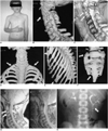
A 12-year-old boy with deformity of the chest wall was admitted to the pediatrics clinic. The physical examination on admission revealed asymmetry in his chest and neck; there was also a sacral dimple and hypoplasia of the right thumb (Fig. 1A). Routine cervical spine films and the postero-anterior (PA) chest film were obtained to search for the etiology of the neck and chest asymmetry. Cervical radiography showed partial vertebral body fusion associated with neural arch fusion of C2-C4 and the wasp-waist sign, and bilateral cervical ribs were noted at the level of C7. The PA chest film revealed rib fusion on the right side at the 1-2 and 8-9 rib level. He underwent axial multislice-helical computed tomography (MCT) with 3-dimensional (3D) reconstructions in order to evaluate the lesions in detail, and also for the differential diagnosis for some of the syndromic disorders that were causing these deformities. These skeletal anomalies were confirmed by 3D-CT (Figs. 1B-D). We also observed partial body fusion of T1-T2 on the 3D sagittal image (Fig. 1E), and this finding was not revealed by plain radiography, CT or the MR images. Spina bifida of L5, lumbalization at the right side of S1 and a sacral curve defect were demonstrated with the 3D-CT scan, and the CT was performed because of the sacral dimple (Fig. 1F). An MRI study was performed to exclude diastematomyelia and syringomyelia. The MRI examination depicted levels of cervical fusion enlargement of the spinal canal and dural sac ectasia (Fig. 1G). The echocardiographic examination revealed mitral valve prolapsus, and the ultrasonographic examination of the abdomen was normal. Intravenous urography (IVU) was performed because of his enuresis nocturna, and it identified a triple renal pelvis in the left kidney (Fig. 1H). Our patient had neither deafness nor ear anomalies.
DISCUSSION
Klippel-Feil syndrome is a complex disorder that consists of congenital fusions of the cervical vertebrae with the patients having a short neck, limitation of the movement of the head or the neck and a low posterior hairline. This syndrome is a result of failure of the normal segmentation of the cervical somites during the 3rd to 8th weeks of gestation (2). Klippel and Feil first reported the clinical features of this syndrome in 1912. In 1919, Feil described additional cases and he distinguished KFS into three different groups according to the degree of involvement (1, 2). Massive fusion of many of the cervical and upper thoracic vertebrae was designated as type I, the fusion at one or two interspaces with occipitoatlantoid fusion, hemivertebrae or other abnormalities in the cervical spine as deemed type II, and last, cervical fusion in combination with lower thoracic or lumbar fusion was deemed as type III (1-3). The morphologic features described in our patient were consistent with type II KFS.
The clinical presentation of KFS is variable. Klippel-Feil syndrome includes a complex of anomalies such as scoliosis (60%), renal anomalies (35%) such as agenesis, dysgenesis, renal ectopia, malrotation, duplication and Sprengel's deformity (30%), deafness (30%), congenital heart disease (14%) such as atrial septal defect and coarctation, cranial and facial asymmetry (13%) and cleft palate (10%) (1, 5). Our patient had additional renal and cardiac anomalies.
The important symptoms and anomalies are usually revealed incidentally on radiological examinations in the mild cases. However, the patients may develop neurological symptoms that are secondary to the degenerative disc disease of the adjacent mobile segments, spinal instability from the hypermobility or from trauma, and spinal stenosis in later decades of life. Therefore, the early diagnosis of KFS is important because of the high risk for other associated diseases in the affected patients (1, 4).
The anterior concave indentation at the site of the absent or reduced interspace between the fused vertebrae is known radiologically as the wasp-waist sign. Nguyen et al. (1) studied cervical spine radiographs of 33 patients with KFS for the patterns of bony fusion and the presence of wasp-waist sign. They reported that the wasp-waist sign was associated with complete vertebral fusion in KFS. In our case, we demonstrated the presence of wasp-waist sign on the 3D CT images.
Thomsen et al. (2) reported that most of the skeletal anomalies are found in type II KFS (2). Our patient was consistent with type II KFS because of the multiple skeletal anomalies without lower thoracic or lumbar fusion.
A high incidence (64%) of genitourinary-tract anomalies that are demonstrated by IVU and on the physical examination are encountered in patients with KFS. The incidence of these anomalies in the three types of the syndrome are essentially same and unilateral renal agenesis is the most common abnormality (3, 6). Our patient had a triple renal pelvis that was shown with IVU. Many kinds of congenital cardiac anomalies are also prominent in this syndrome, and ventricular septal defect is the most commonly encountered lesion (7). Our patient had prolapse of the mitral valve. To the best of our knowledge, only one case has been reported in the literature showing prolapse of the mitral valve associated with KFS (8).
CT scans can delineate fine skeletal structures in great detail with its ability for multislice scanning, and CT may assume a new role for this purpose because of its superior 3D spatial resolution (9). Nowadays, volume rendering is the preferred technique for the musculoskeletal system examinations, and it can improve visualization and better demonstrate subtle findings. This situation may alter the medical management in a significant number of cases (9). Improvements in software have made it quicker and easier to produce high quality images. The utilization of the spiral technique has allowed rapid procurement of overlapping slices for 3D reconstruction of the data without any additional radiation exposure to the patients (10). The disadvantages of this technique are the additional costs associated with the necessary software and the time needed to perform satisfactory reconstructions on the workstation. We found that 3D CT clearly showed the skeletal abnormalities and it helped us make a clear and easy diagnosis in our case. Therefore in our opinion, the diagnostic value of using multislice CT in the cases with skeletal anomalies such as KFS overcome the disadvantages of this technique.
In conclusion, KFS is a clinical and radiological entity with numerous associated anomalies and a variety of complications. The pattern and locations of the fusions provide the diagnostic guidelines for the radiological differential diagnosis. We believe that 3D-CT is helpful in the diagnosis of KFS, and especially in the atypical clinical cases, for delineating the skeletal abnormalities associated with KFS.
 XML Download