

 PDF
PDF ePub
ePub Citation
 Print
Print


The first successful case of living donor liver transplantation (LDLT) was performed by Raia et al. in 1989 (1). In spite of its short history, the difficulties associated with cadaveric donations and serious donor shortages have led to the development and popularization of LDLT (2, 3). In contrast to the other types of major abdominal surgery, LDLT is associated with several specific situations in which arterial bleeding is likely to occur. In effect, LDLT generates a liver graft with a cut surface of variable size and multiple anastomotic sites that are potential sources of bleeding after revascularization (4). In addition, in spite of the refinements that have been made in the medical and surgical techniques, all liver transplantation recipients have some degree of coagulopathy, especially during the perioperative period, as a result of their underlying liver disease (4-6).
Although its incidence and mortality rate have not been precisely determined, arterial bleeding after liver transplantation has sporadically been reported to be an obvious cause of death (4-15). To our knowledge, there has been little published data about post-transplant hemorrhage, its diagnosis with angiography and subsequent treatment with embolization. It seems that most cases of arterial bleeding following liver transplantation have been treated primarily by repeat surgery (4, 6).
At our institution, transcatheter arterial embolization (TAE) has been the first step in the control of presumptive post-transplant arterial bleeding for the past several years. The aims of this retrospective study are to analyze the etiology of arterial bleeding foci in LDLT recipients and to evaluate the efficacy of TAE.
MATERIALS AND METHODS
Patient Population
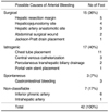
Between April 1999 and March 2001, 32 (26 men and 6 women; age range, 7-62 years; mean age, 43 years) of 195 patients who had undergone LDLT underwent 42 sessions of conventional arteriography in search of bleeding foci of arterial origin within 6 months of the operation. Arterial bleeding was suspected clinically if the symptoms or signs were as follows: abnormally increased drainage of fresh blood through the Jackson-Pratt (JP) tube in 26 sessions, hematochezia or melena in 8 sessions, hemothorax or hemoptysis in 5 sessions, hemobilia in 2 sessions, or an acute decrease in the hemoglobin level in one session. Six patients underwent two or three sessions of arteriography or TAE, because of recurrent arterial bleeding. However, the bleeding foci were different in each of the sessions conducted for the same patient. In all patients, the arterial bleeding occurred within two months (mean, 14 days; range, 1-56 days) after LDLT. Arteriography was performed within three days (mean, 1 day) after the detection of arterial bleeding, depending on the patient's condition.
The underlying causes of hepatic failure and LDLT in these 32 patients were hepatitis B-related liver cirrhosis with or without hepatocellular carcinoma (n=29), secondary biliary cirrhosis due to intrahepatic stones (n=1), fatal fulminant hepatitis due to Wilson's disease (n=1), and biliary atresia (n=1). The grafts involved in the LDLT included the right lobe (n=21), left lobe (n=8), dual left lobe (n=2), and left lateral segment (n=1).
Conventional Arteriography and Transcatheter Arterial Embolization
In all patients, informed consent was obtained from the patient or the patient's family prior to arteriography. Once the right or left femoral artery was punctured, a 5-F end-hole catheter was introduced over a 0.035-inch guide wire (Terumo; Radiofocus, Tokyo, Japan). Following the superior mesenteric and common hepatic arteriographies, which were performed to evaluate the patency of the portal and hepatic arterial anastomoses and potential bleeding foci, the suspected arteries destined for selective arteriography were selected. A provisional identification of the bleeding arteries was made based on the patient's symptoms or signs, radiological findings, including dynamic CT and RBC scans, and the surgical information obtained during LDLT.
In the patients with abnormally increased drainage of fresh blood through the JP tube, selective arteriographies were performed of several specific arteries, for which the arterial bleeding might be stagnant in the JP tube placed area. In those patients with hematochezia or melena, inferior mesenteric or left gastric arteriography was performed following superior mesenteric and common hepatic arteriographies, according to the available clinical information. In those patients with hemothorax or hemoptysis, selective arteriographies of the inferior phrenic artery, intercostal arteries adjacent to the pleural drain tubes, subclavian artery and/or bronchial artery were performed according to the available clinical information. Aortography for the purpose of gaining an understanding of the arterial anatomy prior to selective arteriography was not routinely performed. However, aortography was performed to rule out the possible of there being any missed bleeding foci, if the selective arteriographies of the presumptive arteries did not demonstrate any active bleeding foci.
If the bleeding foci were present on the arteriograms, then either TAE or surgery was undertaken to control the bleeding, subject to the mutual agreement of the surgeons and interventional radiologists. TAE was intended to be the primary treatment, however, surgical management was considered as the primary treatment if the bleeding foci were technically too difficult or dangerous to be embolized, such as in the case of a hepatic arterial anastomotic site or hepatic resection margin.
For TAE, the bleeding arteries were superselected using a 3F-microcatheter (Microferret; Cook, Bjaerverskov, Denmark) and embolized with 0.018-inch-Hilal microcoils (Cook, Bloomington, U.S.A.) or 0.018-inch-Tornado microcoils (Cook, Bloomington, U.S.A.). Gelatin sponge particles (Gelfoam; Upjohn, Kalamazoo, MI) were also used in the case of several specific arteries (intercostal, inferior phrenic and epigastric arteries) prior to embolization using microcoils, to minimize any rebleeding from collateral vessels.
If the bleeding foci were absent on the selective arteriograms and aortogram, explorative laparotomy or clinical observation was chosen by the surgeons, depending on the patient's condition.
Analysis
In all patients, a complete retrospective review of the medical and surgical records and radiological imaging were performed. The following items were documented retrospectively: the presumptive causes and locations of arterial bleeding, the technical and clinical success rates of TAE, and the complications.
A bleeding focus was defined as an extravasation of the contrast media or a pseudoaneurysm that was a likely cause of bleeding. The arterial bleeding was divided into four categories according to the possible related causes: Surgical bleeding was defined as an arterial bleeding related to the LDLT itself and included bleeding from the hepatic resection margin, hepatic arterial anastomotic site, incision wounds, and the hepaticojejunostomy site. Iatrogenic bleeding was defined as bleeding associated with percutaneous invasive procedures, such as transhepatic biliary drainage, central venous access and chest tube insertion. Spontaneous bleeding was defined as bleeding that developed spontaneously, such as gastrointestinal bleeding remote from the hepaticojejunostomy site. Non-classifiable bleeding was defined as bleeding that was not able to be classified properly.
The technical success of TAE was defined as the complete disappearance of arterial bleeding following TAE. Technically impossible or incomplete embolization was regarded as technical failure. Clinical success was defined as an amelioration of the presenting signs or symptoms of arterial bleeding following TAE. Major complications were defined as those necessitating an increased level of care, surgery, prolonged hospital stay, permanent adverse sequelae or death. All other complications were defined as minor complications.
RESULTS
Causes and Locations of Arterial Bleeding
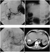
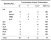
Conventional arteriography demonstrated 42 bleeding foci of arterial origin in 30 sessions (23 patients). The number of bleeding foci detected were one in 22 sessions (15 patients), two in six sessions (6 patients), and more than three in two sessions (2 patients). The distribution of the bleeding foci is shown in Table 1. The bleeding foci originated most frequently from the hepatic artery (14/42, 33%) and intercostal artery (11/42, 26%).
Based on a complete review of the clinical, interventional and surgical data, the bleeding foci were classified into 4 categories corresponding to the most likely causes of arterial bleeding (Table 2). In this way, it was demonstrated that, in addition to surgical bleeding (36%), iatrogenic bleeding (40%) was also a major cause of arterial bleeding after LDLT.
Transcatheter Arterial Embolization
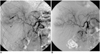
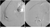
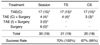
TAE was technically successful in 33 of the 42 foci of active arterial bleeding (Table 1) (Figs. 1, 2). However, TAE failed in the remaining 9 foci of active arterial bleeding, due to technically difficult superselection of the corresponding bleeding artery or the potential high risk associated with TAE (Fig. 3). These bleeding foci consisted of hepatic artery anastomotic sites (n=2), hepatic resection margins (n=4) and hepaticojejunostomies (n=3), and they were treated by surgery.
Embolization was completed in 21 sessions (Table 3). However, in three sessions, additional immediate surgery had to be conducted following successful TAE for the evacuation of hematoma. However, there was no evidence of active arterial bleeding in the operation field in any of these cases. In 20 of the 21 sessions, the patients showed a clinical improvement in the active arterial bleeding after successful TAE. One patient did not show any clinical improvement, because of recurrent arterial bleeding of the same focus. This patient had been treated by anti-coagulation therapy due to cerebral infarction, resulting in a severe iatrogenic bleeding tendency, and died of multifocal hemorrhage including cerebral hemorrhage several days after TAE. The overall technical and clinical success rates for the 30 sessions of TAE were 21 (70%) and 20 (67%), respectively. These relatively low success rates can be ascribed to the three bleeding foci (hepatic resection margins, hepatic artery anastomotic sites and hepaticojejunostomy sites), in which embolization was either incomplete or impossible. In the case of the three bleeding foci, the technical success rate was only 2 (18%) out of 11 sessions. If these bleeding foci were excluded, the overall technical and clinical success rates were 19 (100%) and 18 (95%) out of 19 sessions.
Six (19%) patients underwent two or three sessions of arteriography, because of recurrent arterial bleeding. However, different bleeding foci were treated in each session of arteriography conducted for the same patient. Recurrent bleeding at the same foci was present in only one patient, as described above.
On the other hand, arteriography showed no active bleeding focus in 12 sessions (11 patients). These cases were managed either by surgery (6 patients) or close clinical observation (5 patients) with transfusion, depending on the patient's condition. In three of the former six patients, surgery allowed the discovery of active arterial bleeding at the hepatic artery anastomotic site (n=1), hepatic resection margin (n=1), and venous oozing (n=1) at the anastomosis of the hepatic vein and vena cava. However, a retrospective review of the selective angiographic images of these patients did not demonstrate the presence of any bleeding focus. In the remaining three patients, surgery did not lead to the discovery of any bleeding focus, and they only underwent hematoma evacuation. After either conservative or surgical management, the signs of active bleeding ameliorated in all 11 patients.
During the 6 month follow-up period after LDLT, there were no major TAE-related complications.
DISCUSSION
According to our results, arterial bleeding after LDLT was not confined to surgical causes. Surgical bleeding was one of the most common causes of bleeding after LDLT, however, iatrogenic bleeding was even more common than surgical bleeding in our retrospective study. Coagulopathy after LDLT is inevitable for a certain period of time in most liver transplant recipients, until the graft's function is normalized. The risk of iatrogenic bleeding is also high in the early post-LDLT period. Thus, clinicians should make a particular effort to prevent iatrogenic bleeding during percutaneous procedures after LDLT.
Some amount of surgical bleeding from the hepatic artery at the hepatic arterial anastomotic site, hepatic resection margin or hepaticojejunostomy site is inevitable, because LDLT generates a liver graft with variable-sized cut surfaces and multiple anastomotic sites. In fact, bleeding from the hepatic artery was the most common cause of bleeding in our study. However, performing TAE in the case of bleeding from the hepatic artery following LDLT was virtually impossible, because of the difficulty involved in the superselection of the bleeding arteries, due to the existence of fine arterial feeders, hepatic arterial anastomotic stenosis, a tortuous arterial course or multifocal occurrences. Furthermore, performing TAE of the hepatic artery runs the risk of hepatic infarction or failure following inadvertent diffuse proximal embolization (16). Marshall et al. (17) reported that three of nine patients with extrahepatic pseudoaneurysm following liver transplantation underwent effective coil embolization, however all patients subsequently underwent retransplantation due to graft ischemia.
Accordingly, we achieved technical success in only four of the 14 foci involving bleeding from the hepatic arterial branches. Although TAE can be performed in some patients with massive bleeding from the hepatic artery, we consider that surgical management is the treatment of choice for the management of surgically induced bleeding from the hepatic artery anastomotic site, hepatic resection margin or hepaticojejunostomy site.
The next most frequently bleeding arteries were the intercostal and inferior phrenic arteries. The cause of bleeding from the intercostal artery was iatrogenic bleeding in most cases, resulting from either placement of a drain tube or central venous catheterization. The cause of bleeding from the inferior phrenic artery was uncertain, however it might have been related to various causes, such as intraoperative retractor injury, chest tube insertion or spontaneous bleeding, and it must be routinely evaluated to rule out active bleeding.
Knowledge of the clinical symptoms or signs of arterial bleeding provided by the clinicians is very helpful prior to the performance of arteriography. A JP drain was the most common clinical clue to the presence of arterial bleeding after LDLT. A lot of arteries are involved in this type of manifestation. Bleeding can occur from the hepatic, intercostal, inferior phrenic or pancreaticoduodenal arteries or from the jejunal branches of the superior mesenteric artery. Unusually, bleeding from the deep circumflex iliac and inferior epigastric arteries was manifested as JP bleeding in one case. Therefore, in our opinion, arteriograms of the inferior phrenic and intercostals arteries, as well as of the superior mesenteric and common hepatic arteries, should be surveyed in cases of JP drain bleeding. In addition, those arteries supplying the lower thoracic and abdominal walls should also be considered as possible sources of arterial bleeding.
As in the case of general surgical patients, LDLT recipients are also subject to bleeding from stress ulceration, peptic ulcer and angiodysplasia of the colon (4, 6, 18). Not uncommonly, this bleeding tends to be resolved only by proper medical treatment (4). Clinical observation may be sufficient whenever the patient's condition tolerates it. In our study, four patients with hematochezia or melena showed negative outcomes on arteriography, but improved clinically without surgery or TAE. In fact, TAE was very effective in cases of positive arteriography, and all four patients with arteriographically proven gastrointestinal bleeding improved after only one session of TAE.
In the conventional arteriogram, a negative outcome does not always guarantee the absence of actual bleeding. In our study, surgery demonstrated active bleeding foci in three of six patients without active bleeding on selective arteriography. This discrepancy seems to have been caused by minor or intermittent arterial bleeding or venous oozing. In contrast, surgery did not lead to the detection of any bleeding focus in the three remaining patients.
In conclusion, percutaneous procedures constituted one of the major causes of post-LDLT arterial bleeding. Therefore, extra caution should be taken in LDLT patients. Technical limitations were encountered when using TAE for the management of hepatic arterial bleeding. However, in the other locations, TAE seems to be effective and safe for the control of arterial bleeding after LDLT.
 XML Download