

 PDF
PDF ePub
ePub Citation
 Print
Print


Primary lymphoma of the urinary bladder accounts for less than 1% of the neoplasms occurring in this organ. A few cases have been reported in the radiology literatures (1-3), but in no instance has been described associated calcification. We report the CT and MR imaging findings in a patient with urinary bladder lymphoma involving internal calcification.
CASE REPORT
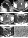
A 30-year-old woman presented with a two-month history of gross hematuria and dysuria. The urine analysis results were 250/ul of RBC and 500/ul of WBC and the serum albumin level was 2.3 g/dl. Intravenous urography revealed a lobulated filling defect and irregular amorphous calcification in the left lateral wall of the urinary bladder, while ultrasonography demonstrated asymmetric hypoechoic diffuse thickening of bladder wall with acoustic shadowing in the left lateral wall suggesting calcification (Fig. 1A). Pelvic CT depicted eccentric lateral wall thickening with calcification and heterogeneous enhancement (Figs. 1B, C). T1-weighted MR image depicted low-signal-intensity eccentric lateral wall thickening, and T2-weighted MR image demonstrated heterogeneous high signal intensity (Figs. 1D, E). There was no evidence of pelvic lymph node involvement. There was a small amount of ascites, but this was probably due to hypoalbuminemia. Their radiologic findings, including those of chest radiography and abdominal CT, indicated no lymph node enlargement or hepatosplenomegaly. Cystoscopy revealed diffuse hyperemic edematous mucosal change, calcification, and mucosal ulceration of the left lateral wall. Ultrasonography-guided biopsy was performed, and histologic examination revealed high-grade peripheral T-cell lymphoma accompanied by focal necrosis (Fig. 1F). Two weeks later, prior to any appropriate treatment, the patient died.
DISCUSSION
Because lymphoid tissues are not normally found in the bladder, a primary lymphoma rarely occurs there (1-4). The tumor commonly affects woman and there is often a history of chronic cystitis, considered important etiologically. It has been suggested that the neoplasm associated with chronic cystitis may originate in the lymphocytes present in the submucosa of the bladder as a result of an inflammatory or immune response (2). The radiologic findings of urinary bladder lymphoma vary, though from a review of the literature, certain patterns emerge. The tumor appears most frequently as a sessile solitary mass (66%); multiple sessile masses are less frequent (14%), and a polypoid mass is occasionally present (10%). Diffuse thickening of the bladder wall may occur, but is infrequent (10%). Despite these patterns of occurrence, there are no characteristic findings which distinguish lymphoma of the urinary bladder from other tumors (1). In our case, ultrasonography and pelvic CT revealed diffuse bladder wall thickening, a finding which can frequently mimic transitional cell carcinoma (TCC) (1, 2).
Pre-treatment calcification in lymphoma patients is extremely rare, and its pathogenesis is uncertain. Generally, tissue calcifications is metastatic or dystrophic (5-7); The former is caused by excessive or unstable calcium ion concentration in the blood. In our case, however, this level was normal, and this explanation was thus less likely. Dystrophic calcification, on the other hand, frequently occurs in degenerated or necrotic tissue (6), conditions which result from infarction and lead to calcification. Such infarction occurs typically in histologically aggressive lymphoma (7). In our case, high grade T-cell lymphoma accompanied by necrosis was diagnosed, and the pathogenesis for calcification was probably a dystrophic calcification.
In TCC and urachal carcinoma, calcification occurs within the bladder wall. Infiltration appears to be more diffuse in lymphoma than in TCC, and in the former, hydronephrosis seems to be uncommon (8). Urachal carcinoma also commonly involves calcification, which typically occurs at the bladder dome. Schistosomiasis may produce mural calcification which shows with a typical thin arcuate pattern, and may also be associated with calcification in other parts of the urinary tract. Other entities that cause bladder wall injury can predispose to dystrophic calcification and include alkaline incrusted cystitis, cytotoxin cystitis, radiation therapy, and tuberculosis (9).
Treatment of primary lymphoma of the urinary bladder is not uniform and the procedures employed have included radical cystectomy, radiation therapy, and chemotherapy. For prognosis, histologic grade is probably a useful indicator, and the overall prognosis appears to be good (2).
In summary, we report a case of primary lymphoma of the urinary bladder associated with dense internal calcification. A case of this nature has not previously been reported in the literature in English.
 XML Download