This article has been
cited by other articles in ScienceCentral.
Abstract
Objective
To evaluate the effects of cyst ablation with absolute ethanol in autosomal-dominant polycystic kidney disease (ADPKD) patients with symptomatic cysts.
Materials and Methods
Using absolute ethanol, cyst ablation was performed in 11 patients with documented ADPKD who suffered cyst pain refractory to medical treatment. An ethanol solution was instilled into the largest symptomatic cysts through a catheter. We assessed the therapeutic efficacy of the procedure by tracking subjective pain relief during a 3 to 24-month follow-up period after ablation.
Results
At follow-up, we found that the duration of subjective pain relief was 12 to 24 months in seven patients, 4 to11 months in one, and less than 3 months in three.
Conclusion
Selective ablation of a symptomatic cyst may be a valid option in managing chronic pain caused by one or a few large cysts in ADPKD patients.
Go to :

Keywords: Kidney, cysts, Kidney, disease, Kidney, interventional procedures, Sclerotherapy
It is well known that the progressive enlargement of renal cysts in autosomal-dominant polycystic kidney disease (ADPKD) patients is frequently associated with abdominal pain, which afflicts about 60% of patients (
1). When medical analgesics fail to alleviate the pain, cyst decompression procedures, including surgical intervention, may be necessary (
1-
4).
Although the ablation of simple cysts has proved effective, the use of this procedure for symptomatic patients with ADPKD has only recently been investigated (
5-
7). Percutaneous sclerotherapy may be difficult in ADPKD, due to the presence of multiple cysts and the difficulty in identifying those which cause pain, selective ablation is, however, effective for the management of chronic pain caused by one or a few large cysts (
5,
6).
The purpose of this study was to evaluate the effects of cyst sclerotherapy with absolute ethanol in ADPKD patients with symptomatic large cyst(s).
MATERIALS AND METHODS
Eleven patients with ADPKD [M:F = 6:5; age, 20-73 (average, 41.8) years] were involved in this study. All complained of abdominal or flank pain caused by enlarged cysts, but none required pain-relieving surgery. Four had hypertension, and two complained of a palpable abdominal mass. A patient with a large and painful cyst who did not respond to medical treatment was considered a candidate for cyst sclerotherapy. To localize the pain, we reviewed a patient's detailed clinical history and decided to target the largest cysts, as determined by radiological imaging (US and/or CT). The procedure was restricted to patients who had severe pain attributed to cysts greater than 5 cm in diameter. In all cases, informed consent was obtained.
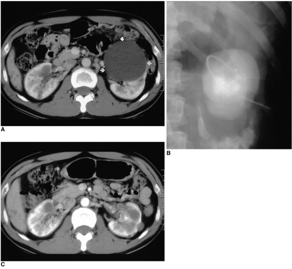
All procedures were performed on an in-patient basis. The patient was placed either in a prone or recumbent position after confirming the location of the largest cyst using US. After sterile preparation, a 22-gauge puncture needle was inserted into the cyst under ultrasonographic (US) guidance. It was then removed, and replaced by a 6-F pigtail catheter, inserted over the guidewire. To determine the existence of any connection with the collecting system and to test for the extravasation of cystic fluid, contrast medium was injected (
Fig. 1), and to permit natural drainage, the catheter was left open. Absolute ethanol (99.8%: J.T. Baker, Holland) was used as the sclerosing agent. On the following day, after completely draining the cystic fluid and ascertaining its volume, we instilled an amount of sclerosing agent equal to one quarter of the volume of the drained fluid. The cystic volume was calculated using the formula: anterior-posterior diameter × transverse diameter × length × 0.523 cc. During this period, the patient was instructed to lie in the supine, prone, and left and right lateral positions successively to facilitate adequate exposure of the cyst wall to the sclerosing agent. The ethanol was then aspirated, and the catheter was left in place for the next treatment session, performed on the following day. The pigtail catheter was removed after completely draining all fluid.
 | Fig. 1
ADPKD in a 34-year-old man.
A. Axial contrast enhanced CT scan depicts multiple cysts in both kidneys. An 8-cm cyst, present in the left kidney (arrows) was ablated.
B. Sinogram obtained during sclerotherapy shows no evidence of extravasation or connection with the collecting system.
C. Twelve-month follow-up CT reveals no evidence of re-expansion of the treated cyst. Pain was relieved for 18 months.

|
The mean follow-up period was 12 (range, 3 to 24) months. In five patients, follow-up CT studies were performed at 9-15 (mean, 11.6) months. Therapeutic efficacy was assessed on the basis of the patients' subjective judgment of their symptoms (abdominal and/or flank pain) during their most recent follow-up visit. Blood pressure was also checked, and any changes were recorded.
Go to :

RESULTS
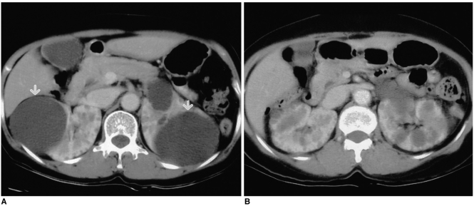
Each patient was treated for the largest cyst on the symptomatic side, and all tolerated the procedure very well. Six of the cysts were on the right side, and four were on the left; in one case, cysts were present bilaterally (
Fig. 2). In all patients, the punctured cysts were 5-10 (mean, 7.3) cm in diameter. The average volume of the sclerosing agent was 30 (range 15-100) cc, and the catheter was kept in place 2 to 5 (mean, 2.9) days. Sclerotherapy gave rise to complications in five patients. Four experienced pain, that was treated conservatively, and one suffered mild and transient gross hematuria.
 | Fig. 2
ADPKD in a 52-year-old woman.
A. Axial contrast-enhanced CT scan obtained prior to sclerotherapy depicts symptomatic cysts in both kidneys (arrows). Cyst ablation was performed bilaterally.
B. Follow-up CT at 18 months shows reduced renal volume and no evidence of cyst re-expansion. Pain relief lasted 18 months.

|
At follow-up, we found that the duration of subjective pain relief was 12 to 24 months in seven patients, 4 to11 months in one, and less than 3 months in three. After the procedure, one of four patients with hypertension became normotensive, and in two with a palpable mass, symptoms improved.
Go to :

DISCUSSION
ADPKD is characterized by progressively enlarging cysts associated with hypertension, renal failure, pain, hematuria and infection (
2). Abdominal discomfort or lumbago is one of the most frequent, as well as difficult to manage, complications of the condition. The acute onset of pain is often attributed to a ruptured cyst or blood vessel, with extravasation of cystic fluid or blood into perinephric tissues (
3).
Chronic pain syndrome is well documented in ADPKD patients with large cysts and/or much-enlarged kidneys (
3,
8). The impression that severe pain occurs under these circumstances is supported by the observation made by Elzinga et al. that 80% of patients who had experienced pain were pain-free one year after surgical decompression of their cysts (
9). Pain occurs due to compression by surrounding tissues, traction on the pedicle of the kidney, and distension of the renal capsule. The severity of pain generally correlates with the size of the kidneys, but there are some exceptions. Pain related directly to cyst formation tends to be steady and nagging discomfort, exacerbated by standing and walking. Patients can frequently localize the source of pain with one finger, anterior abdominal being more common than localized back pain. A detailed history focusing on pain location and duration, and associated symptoms and maneuvers to produce relief, will frequently distinguish pain related directly to cyst formation from mechanical back pain due to changes in posture caused by enlarging cysts (
1). For this reason we obtained a detailed clinical history and decided to target the largest cysts depicted at radiological imaging (US and/or CT).
When medical analgesics fail to relieve intolerable pain, cyst decompression procedures can be effective (
10). An early technique employed was percutaneous aspiration of the cysts under ultrasonographic guidance (
11). Simple cyst aspiration can relieve pain temporarily, supporting the hypothesis that cyst distention can cause pain (
10), but aspiration alone is inefficient because cysts will reaccumulate fluid due to an active chloride transport process (
1). Thus, cyst contraction involving the use of a sclerosing agent may be more effective in lessening the symptoms associated with simple renal cysts as well as those occurring in ADPKD. For treating extremely large kidneys with multiple large cysts, cyst decortication and even nephrectomy have been effectively performed using laparoscopic techniques (
12).
Percutaneous sclerotherapy is an effective method for treating benign renal cysts (
13-
15). Several agents have been used to destroy the cyst wall epithelium, including ethanol (
13-
16), minocycline hydrochloride (
6,
7), povidone-iodine (
17), acetic acid (
18), and ethanolamine oleate (
19); of these various sclerosants, absolute ethanol was used in the present study. This rapidly inactivates the secreting cells of the cysts, and because it is very slow to penetrate the fibrous cystic capsule, can be removed before the renal parenchyma is affected (
14). Seo et al., however, found that in patients treated with large amounts, its systemic absorption led to fever and a drunken state (
18). Because the volume of the sclerosing agent used was about one quarter that of the cystic volume, there were no such cases in the present study; mild pain and transient hematuria were the only complications. Recurrence after alcohol sclerotherapy was reported by Seo et al. in over 30% of cases (
18), and was probably caused by incomplete ablation of the cystic wall due to dilution by the fluid remaining in the cyst (
2). Repeated injections of sclerosing agent have led to substantially decreased rates of recurrence (
14,
20). Thus, in order to facilitate multiple treatment sessions, we routinely inserted a catheter into a cyst. A small-caliber pigtail catheter was used, and there was no damage to the collecting system or adjacent organs. Thus, sclerotherapy is a safe procedure, and the reaccumulation of cystic fluid is reduced by the repeated instillation of a sclerosing agent through the catheter.
However, the application of this ablation technique is not easy in symptomatic ADPKD patients with many cysts into which catheters should be inserted. Kim et al. (
5) recently reported that percutaneous needle aspiration and the intracystic injection of a mixture of n-butyl cyanoacrylate with iodized oil, performed during the same treatment session, is effective for the ablation of multiple renal cysts.
Our results showed that the percentage probability of being pain free one year after sclerotherapy was 64%. Thus, the selective ablation of symptomatic cysts present in ADPKD patients should be considered a valid option in managing chronic pain caused by one or more large cysts.
To assess the long-term effects of these procedures on the outcome of ADPKD in terms of the duration of pain relief, further study and follow-up is needed.
Go to :


 PDF
PDF ePub
ePub Citation
 Print
Print


 XML Download