Abstract
We report the hemodynamic assessment in a patient with cerebral arteriovenous malformation using time-resolved magnetic resonance angiography (TR-MRA), a non-invasive modality, and catheter-based digital subtraction angiography (DSA), before and after embolization. Comparison of the results showed that TR-MRA produced very fast dynamic images and the findings closely matched those obtained at DSA. For initial work-up and follow-up studies in patients with vascular lesions, TR-MRA and DSA are therefore comparable.
Conventional catheter-based digital subtraction angiography (DSA) is a gold-standard technique used to diagnose cerebral arteriovenous malformation (AVM) and also where embolization is required. The procedures involved are, however, invasive, requiring hospitalization, careful manipulation during the study itself, and large amounts of both contrast medium and radiation. In addition, complications such as embolism or dissection may occur during or after the procedure. For the diagnosis and evaluation of cerebral vascular lesions, less invasive methods, including several MR angiographic techniques, have been introduced, and among these, both the 3-D phase-contrast and contrast-enhanced 3-D time-of-flight technique are now widely employed. These clearly depict the anatomic features of cerebral AVM, but give only static information (1, 2). To predict the effectiveness of treatment after endovascular management or radiosurgery in cases involving vascular-based lesions such as AVM, the early recognition of hemodynamic change is, however, important (3), and to this end, time-resolved magnetic resonance angiography (TR-MRA) with contrast bolus injection has recently been introduced. The technique was introduced and refined owing to the development of gradient systems for ultra-fast imaging after injection of a contrast agent bolus (4, 5). Nowadays, TR-MRA can be used to obtain fast, dynamic, contrast-enhanced vascular images of selected sections at a temporal resolution of 300 - 400msec per frame. It is a subsecond technique which permits direct observation of the fast hemodynamic changes occurring in normal or abnormal vascular alterations such as AVM or other lesions that contain blood pool space.
In this paper we describe and assess a new modality, TR-MRA, used for the evaluation of AVM after embolization, and compare the findings with those of DSA.
A 36-year-old man with sudden seizure attended our hospital. Initial physical examination detected no neurologic symptoms, but electroencephalography revealed partial seizure in the left temporal region.
For MR imaging, a 1.5-T whole-body unit (Vision; Siemens, Erlangen, Germany) with a standard gradient system (25 mT/m) was employed, and a standard circularly polarized head coil was used for radio frequency transmission and detection. Prior to imaging, an 18-gauge IV catheter was inserted in the right antecubital vein and a standard clinical dose of Gd-DTPA (15ml at 3ml/s following the injection of 12ml normal saline at 3ml/s) was automatically injected. T1-, T2- and enhanced T1-weighted images were obtained (TR/TE=600/14 for T1WI and enhanced T1WI; TR/TE/acquisition = 4500/120/2 for TSE T2WI). TR-MRA was performed twice, before and just after embolization, using a snapshot FLASH sequence optimized for 2-D projection imaging, and the parameters were as follows: minimum TR/TE, 4.2/1.5 ms; slab thickness, 45 mm; flip angle, 45°; field of view (FOV), 220 mm; matrix, 256×256. To increase temporal resolution, the FOV employed was rectangular, and to assess hemodynamic change during an uninterrupted 34-second period, 102 coronally directed TR-MRA images were obtained using one slice frame.
Using a Multistar T.O.P. unit (Siemens) and a standard anteroposterior and lateral projection format (matrix, 1024×1024; 3 frames per second), DSA was performed via the right femoral artery before and immediately after embolization. Both the internal carotid and vertebral arteries were selectively catheterized using conventional methods.
After diagnostic angiography, a Microferret-18 microcatheter (Cook, Bloomington, Ind., U.S.A.) was coaxially introduced into feeding arteries through a 5-F guiding catheter placed in the proximal left internal carotid artery. For embolization, 5-0 silk suture cut into 0.5- to 1-cm lengths was hand-loaded into the microcatheter hub, and saline solution contained in a 1-ml Luer-Lok syringe was injected. The microcatheter was flushed after the delivery of each piece of silk, and to assess the catheter's position and the rate of blood flow through the vascular pedicle being embolized, fluoroscopy was used intermittently.
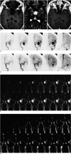
T1- and T2-weighted images depicted a wedge-shaped lesion containing multiple, variable-sized, linear or round signal void structures in the left temporal lobe, while Gd-DTPA-enhanced T1-weighted images showed minimal enhancement (Figs. 1A-C). Before embolization, DSA demonstrated a large AVM nidus fed from a branch of the left middle cerebral artery, and early drainage via a dilated vein, mainly to the distal left sigmoid sinus, was observed. There was early visualization of the nidus and left internal jugular vein, and at delayed imaging, decreased opacity of contrast material in the unaffected part of brain was noted. After embolization of the AVM nidus, visualization of the previously noted dilated vein was delayed, and contrast filling of the unaffected part of brain was almost normal, as was the appearance of the left internal jugular vein (Figs. 1D, E). The inferomedial part of the nidus still remained after embolization, but visualization was delayed more than before.
At TR-MRA prior to embolization, there was, as at DSA, early visualization of the AVM nidus, left internal jugular vein, and dilated draining vein. Visualization of the weakly enhanced superior sagittal sinus, on the other hand, was delayed, suggesting delayed blood flow and decreased blood volume compared to normal parenchyma. At TR-MRA performed immediately after embolization, visualization of the dilated draining vein, and the remaining small part of the nidus, located inferomedially, was delayed. Filling of the superior sagittal sinus and left internal jugular vein was normal, however, suggesting the regulation of blood flow to normal brain parenchyma but decreased blood flow to the AVM (Figs. 1F, G).
Both before and after embolization, DSA and TR-MRA demonstrated similar patterns of rapid hemodynamic contrast filling of the AVM and remaining cerebral parenchyma.
Due to operational ease and reduced risk, contrast-enhanced MR angiography is now more widely used than DSA, and the results obtained are of high quality. Time-resolved MRA, introduced and refined due to the development of gradient systems for ultra-fast imaging after injection of a contrast agent bolus, can obtain consecutive images with a temporal resolution of 300-400 msec per frame. The technique can be used to assess fast hemodynamic change occurring in normal or abnormal vasculature, and is used in cases involving cerebral AVM or other lesions that contain blood pool space. As the gross pathologic appearance of an AVM is essentially a tangled cluster of dilated, tortuous vessels resulting from the preservation of primitive direct communication between arterial and venous channels without an intervening capillary bed, TR-MRA, like DSA, can be used to assess the fast hemodynamic change mentioned above and to determine the effectiveness of surgery, or after embolization, as in our case.
Complete surgical resection is generally considered the treatment of choice for cerebral AVM, and the utility of preoperative embolization in patients who are to undergo this treatment has been described in published reports (6, 7). The benefits involved include shorter operating time, decreased blood loss, and control of deep inaccessible blood vessels. If a cerebral AVM is deeply located, radiosurgery is another possible choice.
The evaluation of post-treatment or adjunctive embolized AVM is important, and even after radiosurgery, early angiography is needed (3). Despite its advantages, however, DSA is an invasive study involving substantial exposure to radiation, and a further drawback is that complications such as embolism or dissection may occur during or after the procedure. MRA, however, is safe, less invasive, less time consuming, and easy to perform without hospitalization. The disadvantage of conventional MRA is that due to poor visualization of vessels, it cannot replace pre-treatment catheter-based DSA (8); TR-MRA, on the other hand, permits assessment of the fast hemodynamic change occurring in cerebral AVM just after the embolization of major branches, as in this study.
TR-MRA can be used before and after embolization of cerebral AVM, in radiotherapy, and postoperatively in the early detection of residual or recurrent cerebral AVM.
In conclusion, hemodynamic assessment of cerebral AVM with both TR-MRA (before and after embolization) and conventional DSA showed that both modalities provided fast hemodynamic images. We suggest that TR-MRA is comparable to catheter-based DSA and could be an alternative for the evaluation of lesions which contain blood pool space, and for follow-up evaluation of such lesions.
Figures and Tables
Fig. 1
Multiple round and linear signal-void structures, which form a wedge-shape defect, are present in the left temporal lobe, suggesting the presence of an AVM in a 36-year-old man.
A, B, C. T1-(A), T2-(B), and enhanced T1-weighted (C) axial MR images are shown (TR/TE = 600/14 for T1WI and enhanced T1WI; TR/TE/acquisition = 4500/120/2 for TSE T2WI). After the injection of contrast medium, enhancement was minimal.
D. Prior to embolization, a large AVM nidus fed from a branch of the left middle cerebral artery is apparent, and there is early drainage, mainly to the distal left sigmoid sinus, via a dilated vein. Note early visualization of the left internal jugular vein. During the capillary phase, decreased opacity of contrast medium in the rest of the cerebral parenchyma was also noted.
E. After embolization with 5-0 silk suture, the previously noted dilated vein has completely disappeared and contrast filling in the remaining part of the brain is almost normal. A small part of the AVM, seen inferomedially, remains, and the filling time and density of the superior sagittal sinus and left internal jugular vein have normalized.
F. TR-MRA acquired prior to embolization shows, as does DSA, early visualization of the AVM nidus and dilated draining vein, and delayed visualization of the weakly enhanced superior sagittal sinus. Additionally, as in DSA, the left internal jugular vein is visualized during the early arterial phase.
G. TR-MRA obtained immediately after embolization shows that visualization of the dilated draining vein and remaining part of the AVM nidus, seen inferomedially, is delayed. Early visualization and normal filling of the superior sagittal sinus is noted, suggesting the regulation of blood flow in the remaining part.

References
1. Huston J III, Rufenacht DA, Ehman RL, Wiebers DO. Intracranial aneurysms and vascular malformations: comparison of time-of-flight and phase-contrast MR angiography. Radiology. 1991. 181:721–730.
2. Marks MP, Pelc MJ, Ross MR, Enzmann DR. Determination of cerebral blood flow with a phase-contrast cine MR imaging technique: evaluation of normal subjects and patients with arteriovenous malformation. Radiology. 1992. 182:467–476.
3. Oppenheim C, Meder JF, Trystram D, et al. Radiosurgery of cerebral arteriovenous malformations: is an early angiogram needed? AJNR Am J Neuroradiol. 1990. 20:475–481.
4. Hennig J, Scheffler K, Laubenberger J, Strecker R. Time-resolved projection angiography after bolus injection of contrast agent. Magn Reson Med. 1997. 37:341–345.
5. Wang Y, Johnston DL, Breen JF, et al. Dynamic MR digital subtraction angiography using contrast enhancement, fast data acquisition, and complex subtraction. Magn Reson Med. 1996. 36:551–556.
6. DeMeritt JS, Pile-Spellman J, Mast H, et al. Outcome analysis of preoperative embolization with N-butyl cyanoacrylate in cerebral arteriovenous malformations. AJNR Am J Neuroradiol. 1995. 16:1801–1807.
7. Jafar JJ, Davis AJ, Berenstein A, Choi IS, Kupersmith MJ. The effect of embolization with N-butyl cyanoacrylate prior to surgical resection of cerebral arteriovenous malformations. J Neurosurg. 1993. 78:60–69.
8. Dobson MJ, Hartley RWJ, Ashleigh R, Watson Y, Hawnaur JM. MR angiography and MR imaging of symptomatic vascular malformations. Clinical Radiology. 1997. 52:595–602.


 PDF
PDF ePub
ePub Citation
 Print
Print


 XML Download