

 PDF
PDF ePub
ePub Citation
 Print
Print


Tailgut cysts, or retrorectal hamartomas, are rare congenital lesions originating from vestiges of the embryonic hindgut (1-4). They are characterized by a variety of intestinal types of epithelium, most often columnar (4). The findings of imaging studies of tailgut cysts, including those using barium enema, US, CT and MRI, have been described (1, 5-7). Most reports stated that they were located in the retrorectal presacral space, and digital rectal examination thus facilitated their detection and diagnosis. Very rarely is this lesion located perirenally. Only one such case, for which imaging findings were unavailable, has been reported: in the patient involved, both a daughter lesion and the main presacral tailgut cyst were found in the perirenal area (2). We describe a case in which a tailgut cyst was located exclusively in the perirenal region, an unusual site, and describe the related imaging findings.
CASE REPORT
A 22-year-old woman was referred for abdominal discomfort and urinary frequency, and at abdominal sonography a huge cyst was detected in the left lower quadrant. The left abdomen contained a palpable mass, but digital rectal examination revealed nothing of significance.
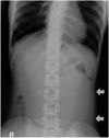
Plain radiography revealed a large soft tissue mass lying in the left abdomen (Fig. 1A), and abdominal CT showed a homogeneous unilocular cystic mass lying anterior to the left kidney, which was compressed and shifted posteriorly by it (Fig. 1B). The mass had a thin, enhancing rim, caused the left ureter to shift laterally (Fig. 1C), and extended to the level of bifurcation of the left common iliac artery (Figs. 1D, E). The patient's serum CA 19-9 level was less than 5U/ml, and CA 125 was 17U/ml, each within the normal range. A benign retroperitoneal cyst such as a lymphangioma or mesenteric cyst was thus the more probable differential diagnosis; a neoplasm originating from the ovary or pancreas was less likely.
Via the left paramadian approach, the mass was surgically excised, and was found to measure 20×13 cm. It ruptured during the procedure, and light yellowish, slightly mucous fluid was aspirated. The gross specimen consisted of a 1-5mm-thick cystic wall, lined by papillary fronds of glandular epithelium with a ciliated border and containing arrays of smooth muscle (Figs. 1F, G). After additional microscopic and immunohistochemical studies, a tailgut cyst was diagnosed.
DISCUSSION
Tailgut cyst, or retrorectal hamartoma, is a congenital lesion believed to originate from the remnant of the tailgut. During its development the embryo possesses a true tail, which reaches its largest diameter at the '35th' day of gestation. The anus develops above the tail on the '56th' day, by which time the latter has completely regressed. Remnants of the tailgut or neurenteric cord may be the origin of tailgut cysts (8), and these thus occur in the anatomic region where tailgut vestiges presumably would reside (2). Most tailgut cysts are located in the presacral retrorectal space, and one report has described a retrorectal tailgut cyst with a daughter lesion in the perirenal area (2). We believe that exophytic lateral growth from the retrorectal space or vestiges of the neurenteric canal adjacent to the left kidney is the possible cause of an unusual perirenal location.
The differential diagnoses of retrorectal masses include a diverse entity of congenital inflammatory, neurogenic, osseous, or other miscellaneous tumors, among which the characteristics of teratoma, epidermoid and dermoid cyst, rectal duplication cyst and anal cyst may be similar to those of tailgut cyst. Epidermoid and dermoid cysts are usually unilocular and are lined by stratified squamous epithelium; the latter have dermal appendages but the former do not. Duplication cysts are also unilocular and are lined by epithelium similar to that of the gastrointestinal and respiratory tracts. The epithelium, often with villi, crypts and glands, simulates normal mucosa of the gut. The main distinctive feature is a well-formed muscular wall overlying two layers of muscular bundles containing a nerve plexus.
Tailgut cysts are usually multicystic or multiloculated, and are lined by a wide variety of epithelia which varies from cyst to cyst, or even within the same cyst, and includes the stratified squamous, transitional, stratified columnar, ciliated pseudostratified columnar, and gastric types. In most cases the cyst contains focal, well-formed smooth muscle fibers; the muscle bundles, however, are often disorganized and are present focally, unlike the well-formed continuous two-layer muscle coat seen in duplication cysts (3). In our case, papillary fronds of glandular epithelium with a ciliated border lined the cystic wall, which also contained arrays of smooth muscle. Focal positive staining for smooth muscle was, in addition, detected immunohistochemically.
At CT, tailgut cyst has been reported as a well-margined retrorectal mass with varying attenuation values. Most cases do not involve calcification, the detection of which may favor the diagnosis of a dermoid cyst, teratoma, or other tumor of osseous origin. For differentiation between this cyst and other malignanct tumors, the presence, at CT, of a smooth margin and the absence of invasion of surrounding structures is also helpful. If however, the locules contain inflammatory or keratinized debris, differentiation between a cyst and a solid mass may be difficult (5, 6). At MRI, tailgut cysts are seen as unilocular and well circumscribed at T1-weighted imaging and homogeneously hyperintense at T2-weighted imaging (1), or have a multilocular honeycomb appearance with thin internal septation (5). The MRI appearance of an epidermal cyst in the retrorectal space has also been described as unilocular (10).
For the evaluation of a retrorectal mass, the ability of MRI to detect calcification is limited, particularly where the exclusion of dermoid cyst or teratoma would be helpful. Using fat suppression techniques, however, MRI can reliably diagnose fatty tumors, and because of high intrinsic tissue contrast, solid components can more easily be excluded (5).
Tailgut cysts most commonly involve middle-aged women, but can occur at any age, including infancy. They may present as an asymptomatic mass during physical examination or at childbirth, and if infected, are often misdiagnosed as pilonidial cyst, anorectal fistula, or recurrent retrorectal abscess. Discomfort while sitting, and rectal bleeding, are common symptoms (3, 6), and malignant change, a rare complication, has occasionally been documented (3). The clinical significance of a tailgut cyst mainly concerns the morbidity that can result if its true nature is not suspected and definite surgery is not undertaken. The potential for infection, the recurrence of perirenal fistulas, and the possibility of malignant transformation emphasize the importance of early complete surgical excision of these lesions (3, 8, 9).
In conclusion, tailgut cysts are rare congenital lesions originating from a remnant of the tailgut. Typically, they occur in the presacral retrorectal space, but are also found, for example, in the perirenal space, where tailgut vestiges presumably would reside.
 XML Download