

 PDF
PDF ePub
ePub Citation
 Print
Print


Choriocarcinoma is a neoplasm of syncytiotrophoblastic origin, of which approximately 50% occur after a molar pregnancy, 25% after a normal pregnancy, and 24% after spontaneous abortion (1, 2). It is a highly vascularized tumor and because of the affinity of trophoblasts for blood vessels, shows rapid hematogeneous metastasis. This is commonly to the lung and vagina, although other sites including the brain, genitourinary and gastrointestinal system have also been reported. When manifestation of metastasis precedes that of the primary lesion, the findings of imaging studies present a diagnostic challenge.
We report two cases of choriocarcinoma which metastasized to the liver in patients with acute and massive abdominal bleeding and characteristic angiographic findings.
CASE REPORTS
Case 1
A 33-year-old Asian woman (gravida 3, para 2) presented with upper abdominal pain and distension, and weight loss of 4kg in one month. Her medical history was unremarkable, though an intrauterine device had been inserted after her last pregnancy, four years earlier.
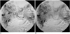
Computed tomography (CT) revealed multiple, irregularly shaped, heterogeneously enhanced mass lesions of variable size in the liver and spleen, suggesting hepatic metastasis from splenic angiosarcoma or multifocal hepatocellular carcinoma. The contour of the uterus appeared to be normal. Diagnostic celiac angiography demonstrated multiple hypervascular masses with abnormal tumor vessels and saccular aneurysmal dilatation of the peripheral end of hepatic arteries at the arterial phase, and persistent visualization of hepatic and splenic vascular lakes at the venous phase, but active bleeding was not apparent (Figs. 1A, B). The size and distribution of the masses were more extensive than CT initially suggested. SMA angiography demonstrated normal portal flow.
Diagnostic laparoscopy was performed, and the findings included multiple hepatic masses, splenomegaly with multiple nodules, and a large (7-8-cm) cystic and solid mass in the left ovary. Laparoscopic biopsy of the hepatic and ovarian masses followed by histolopathologic examination revealed metastatic choriocarcinoma of the liver and a hemorrhagic lutein cyst in the left ovary. The serum beta hCG concentration was very high, at 2,000,000 mIU/mL, but chest radiography revealed nothing abnormal.
Five days after admission, sudden changes in the laboratory data (hemoglobin, 8.7 mg/dl; hematocrit, 26.3%; platelet count, 80,000/mm3) suggested active bleeding, and exploratory laparotomy revealed 4000 cc of blood in the peritoneal cavity and active bleeding from the liver, spleen and both ovaries. Despite bilateral oophorectomy and surgery known as the Pringle method to control bleeding from the liver, the flow could not be staunched and emergency angiography was again performed. Celiac angiography demonstrated massive extravasation of contrast media from the posterior superior and inferior segmental branches of the right hepatic artery and lateral segmental branch of the left hepatic artery, as well as from the branch in the upper pole of the spleen, and immediate embolization was successfully performed using 16 Tornado microcoils (two, 4 mm × 2 cm in size; four, 3 mm × 2 cm; five, 5 mm × 2 cm; five, 6 mm × 2 cm) and gelfoam particles after superselection of the segmental arteries of both hepatic lobes and the spleen. Post-embolization angiography demonstrated no further extravasation of contrast media and intact portal flow. The patient was then observed for evidence of further bleeding.
AST and ALT levels continuously and rapidly increased, however, suggesting progression to hepatic failure and acidosis, and follow-up CT performed 13 days after embolization showed an extensive area of low attenuation with a minimally enhanced region in the enlarged liver and spleen, representing massive hepatic infarction and necrosis. Although the exact cause of this infarction was unknown, it was assumed that embolization was the probable cause of hepatic failure, though some normal parenchymal portions and intact portal flow remained. However, the patient's condition continued to deteriorate, and she died 22 days after embolization.
Case 2
A 33-year-old Asian woman (gravida 4, para 0) was hospitalized via the emergency department for severe abdominal pain without vaginal bleeding. A hydatidiform mole with pulmonary metastasis had been diagnosed about five years earlier and the patient had subsequently undergone eleven cycles of chemotherapy. One year before hospitalization, however, she was lost to follow-up.
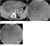
A urinary pregnancy test was positive, showing a serum beta hCG level of over 2,800,000 mIU/mL. Chest CT demonstrated that multiple hematogenous metastatic nodules extended throughout both lungs, while abdominal/pelvic CT revealed huge, ill-defined, heterogeneously enhanced masses in both lobes of the liver and thrombosis in the left portal vein (Fig. 2A). At precontrast CT, somewhat highly attenuated ascites, suggestive of hemoperitoneum, was apparent. The contour of the uterus was normal. The differential diagnoses included other metastatic diseases, primary hepatocellular carcinoma, and other hypervascular hepatic tumors. After the confirmation of laboratory data indicating massive intraperitoneal hemorrhage, exploratory laparotomy was performed.
During surgery, 1500 cc of blood was evacuated from the peritoneum, and the liver was found to have been largely replaced by numerous friable tumor deposits. Several of these in the left lobe were, moreover, ruptured, and were bleeding profusely and continuously. The hepatic masses were ligated, but hemostasis was not achieved.
Two days after surgery a sudden overnight drop in the hemoglobin level, from 9.5 g to 5.5 g, occurred, and the patient's condition continued to deteriorate. A second laparotomy was undertaken to investigate the reason for this drop, and during surgery severe hemorrhage from the friable liver tumors was observed. Hemostasis could not, however, be attained.
On the same day, emergency angiography was performed. Hepatic arterial-phase arteriography demonstrated the presence of huge, multiple, exophytic hypervascular masses with abnormal tumor vessels, saccular aneurysmal dilatation of the hepatic arteries, and arteriovenous shunts, and at the venous phase, persistent bilobar vascular lakes were observed. Active bleeding, was not apparent (Figs. 2B, C). SMA portography revealed thrombosis of the left portal vein, and using gelfoam particles, superselective embolization of the right and left hepatic arteries was successfully performed. Post-embolization angiography demonstrated residual tumor staining supplied by the cystic artery and medial segmental branch of the left hepatic artery, but the previously noted staining of other masses was not observed. The laboratory data also indicated that bleeding has ceased.
Despite the treatment described, the patient's condition continued to deteriorate rapidly. Respiratory failure ensued, and she died 17 days after admission to hospital.
DISCUSSION
Choriocarcinoma is a rare malignant trophoblastic tumor complicating between 1:13,000 and 1:50,000 pregnancies in Caucasian populations and a higher proportion among Asians (3). It is hypervascular, metastasizing hematogenously and producing a dramatic rise in beta hCG levels. The majority of metastases affect the lung (75%), vagina (50%), ovaries, brain, ureter and bowel. Hepatic involvement is not common, affecting only ten percent of patients, and occurring late in the course of the disease (1). Regression of the primary tumor after it has metastasized is not uncommon, and one-third of cases manifest as complications of metastatic disease. Symptoms depend on the location of the metastatic deposit (4, 5). Because of the highly vascular nature of a metastatic lesion similar to a primary tumor, tumoral hemorrhage, whether spontaneous or post-bioptic, can cause great concern. In fact, our first case involving multiple hypervascular hepatic masses in a woman of childbearing age without a history of pregnancy or uterine abnormalities presented with hemoperitoneum and severe anemia. Our initial diagnosis was either hepatic metastasis from splenic angiosarcoma or multifocal hepatocellular carcinoma. Diagnostic laparoscopic biopsy was subsequently performed, resulting in massive hemoperitoneum. In women of childbearing age who present with hepatic masses, the possibility of choriocarcinoma should therefore be emphasized. Because of the risk of hemorrhage, liver biopsy should be deferred until the blood serum beta hCG level is measured, especially if typical angiographic findings of choriocarcinoma are demonstrated.
The angiographic findings for metastatic choriocarcinoma of the brain (6, 7) and kidney (8) are reported in the literature and include prominent hypervascularity, arteriovenous shunt, oncocytic or fusiform aneurysm, and thrombosis. To date, however, liver metastasis has been poorly documented. In our cases, the characteristic angiographic findings for metastatic choriocarcinoma of the liver, as compared with other hypervascular hepatic masses, were a hypervascular mass with aneurysmal dilatation of the peripheral end of the hepatic arteries (grape-lke appearance) at the arterial phase, and persistent vascular lakes at the venous phase. These findings are similar to those of hemangioma, but unlike metastatic choriocarcinoma of the liver, hemangioma has unique CT findings of peripheral enhancement with centripetal flow. In addition, a feature which permits differentiation between metastatic choriocarcinoma and hepatoma is that in the latter, vascular lakes retain contrast media for less time than in the former.
In conclusion, the clinical and imaging presentation of primary choriocarcinoma can be absent or subtle, possibly due to spontaneous regression of the tumor itself, and only a metastatic lesion presents as either a hypervascular liver mass at imaging studies, or progressive anemia or intra-abdominal hemorrhage, clinically. Moreover, CT depicted heterogeneous, multiple enhancing hepatic masses that cannot be easily differentiated from other hypervascular hepatic tumors. Thus, the unique angiographic findings of metastatic choriocarcinoma of the liver, including aneurysmal dilatation of the peripheral hepatic arteries at the arterial phase and persistent vascular lakes at the venous phase, can be helpful for differentiation from other hypervascular hepatic masses.
 XML Download