

 PDF
PDF ePub
ePub Citation
 Print
Print


Castleman's disease was first described as a pathologic entity in 1954 and later defined by Castleman et al. (1) in 1956. A rare lymphoproliferative disorder of unknown etiology and pathogenesis, it most commonly occurs in the mediastinum but also in other areas of the body where lymph nodes are normally found. An axillary location has been reported in only 2% of localized cases (2). We report a case of localized axillary Castleman's disease, describing the imaging features at gray-scale US, power Doppler US, and contrast-enhanced dynamic computed tomography (CT).
CASE REPORT
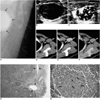
An axillary mass was detected incidentally in a healthy 45-year-old woman who eight months earlier had undergone transabdominal hysterectomy and bilateral salpingo-oophorectomy due to uterine myoma. After surgery she had taken oral estrogen, and breast mammography demonstrated heterogeneous density. Mediolateral oblique imaging depicted a well-defined ovoid mass, 3 cm in size, in the left axilla (Fig. 1A). Gray-scale US using a broad band linear probe (5-12 MHz) revealed a well-defined, uniformly hypoechoic, ovoid axillary mass, 3×2×3 cm in size and with good through transmission (Fig. 1B). Power Doppler US of the mass showed prominent linear and branching peripheral blood flow (Fig. 1C). Precontrast, early phase, and delayed phase spiral CT scanning was performed prior to, 35 seconds after, and 180 seconds after, respectively, initiation of the intravenous infusion of 130 ml of nonionic contrast media at a rate of 2.0 mL/sec. Scanning parameters were 5-mm collimation, 9-mm table feed, and 7-mm reconstruction. At precontrast scanning, the mass was isodense to chest wall muscle (Fig. 1D), while postcontrast scans demonstrated rapid homogeneous enhancement and washout (Figs. 1E and 1F). The mean density of the mass was 48 HU at precontrast scanning, 124 HU at the early phase, and 78 HU at the delayed phase. CT depicted no lymph node enlargement in the mediastinum or abdomen. For US-guided biopsy, a 14-G automated gun was used: microscopic examination revealed tight concentric layering of lymphocytes at the periphery of the lymphoid follicles, with penetration by small capillaries, findings which were pathologically consistent with those of typical hyaline-vascular type Castleman's disease. Surgical excision was performed two weeks after biopsy, and followed by microscopic examination. This showed that in the peripheral portion of the lymph node, the number and size of blood vessels were greater than in the central portion (Fig. 1G), findings which correlated with those of power Doppler US. In the germinal center, multinucleated and pleomorphic follicular dendritic cells were present (Fig. 1H), indicating follicular dendritic cell dysplasia.
DISCUSSION
Since Castleman et al. (1) described 13 cases with the characteristic histopathologic features of a localized mediastinal lymph node hyperplasia in 1956, many such cases have been reported under various names: lymphonodal hamartoma, follicular lymphoreticuloma, angiofollicular lymph node hyperplasia, angiomatous lymphoid hamartoma, and giant lymph node hyperplasia. The condition is currently classified into two major clinical subgroups: localized and disseminated Castleman's disease (3), and two major histological types: hyaline-vascular and plasma-cell (4). Keller et al. (4) found that 91% of all cases were the hyaline-vascular type and 9% were the plasma-cell type, while Weisenburger et al. (5) reported that most cases of disseminated Castleman's disease were the plasma-cell type and in affected patients the prognosis was typically much worse than in those with localized disease.
Although inflammatory and immunological processes are thought to be implicated, the exact etiology of localized Castleman's disease is unknown. It is thought to represent either reactive lymphoid hyperplasia due to chronic antigenic stimulation by a virus, or a developmental growth disturbance (3, 6). In our case, the patient presented no symptoms and the axillary mass was detected incidentally.
In Castleman's disease, gray-scale US reveals a uniform hypoechoic mass (7), while at Doppler US, one that is hypervascular is depicted (8). When differentiating between benign and malignant axillary nodes, shape and intranodal blood flow pattern are important. In a study by Yang et al. (9), gray-scale US showed that the mean longitudinal-transverse axis ratio (±SD) was significantly lower in malignant (1.8 ± 0.6) than in benign nodes (2.6 ± 0.8), while Doppler US demonstrated that both the total number of vessels, and the number of those that were peripheral, were greater in malignant than in benign axillary nodes. In our case, the lymph node showed a low mean longitudinal-transverse axis ratio, 1.5, and pathologic examination indicated that peripherally, many more and larger blood vessels were present, findings which suggested malignancy. Because mammography and US revealed no apparent abnormality, we believed that metastasis from another organ might have occurred. Lymphoid follicular dendritic cell dysplasia, which can become malignant, was diagnosed, and the observed peripheral hypervascularity may be related to this condition. Reactive lymph nodes tend to involve a diffuse histological process and are more likely to preserve a normal vascular pattern, with central hilar vessels (10). On the other hand, malignant changes caused by infiltrating tumor cells lead to the distortion and destruction of preexisting nodal vascular structures. After central malignant infiltration of the lymph node, US-histopathologic correlation of the findings of lymphadenopathy demonstrated the presence of residual subcapsular vessels (10).
At CT, Castleman's disease appears as a homogeneous enhancing mass (7). Our patient underwent triphasic CT for evaluation of the hemodynamics of the lymph node, and postcontrast scanning demonstrated early rapid enhancement and washout. Because rapid homogeneous enhancement of the node occurred, CT did not demonstrate peripheral hypervascularity.
In conclusion, the imaging findings of Castleman's disease localized in the axilla were peripheral hypervascularity at power Doppler US and early rapid enhancement at dynamic CT.
 XML Download