Abstract
The radiologic and clinical findings of foreign bodies in the chest of children are well recognized. Foreign bodies in adults are infrequent, however, and the radiologic findings of these unusual circumstances have rarely been described. We classified various thoracic foreign bodies into three types according to their cause: Type I, Aspiration, Type II, Trauma or Accident; Type III, Iatrogenic. This pictorial essay will illustrate the radiologic findings and consequences of thoracic foreign bodies in adults, which have rarely been described in the radiologic literature. The clinical significance of thoracic foreign bodies will be also be discussed.
The radiologic and clinical findings of foreign bodies in the chest of children are well recognized. Foreign bodies are infrequently found in adults, however, and the radiologic findings of these unusual circumstances have rarely been described. The causes of foreign bodies in adults are much more frequently iatrogenic or traumatic than is the case in children, and where the location is thoracic, the various nonspecific or unusual imaging features occasionally encountered may cause diagnostic problems. A familiarity with the radiologic features of these unusual circumstances can thus be helpful in the early and correct diagnosis of intrathoracic foreign bodies and the judicious management of the patients concerned. In this pictorial essay, we classify various thoracic foreign bodies into three types according to their causes, and illustrate radiologic findings and consequences of thoracic foreign bodies in adults.
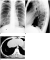
It is well known that foreign body aspiration is more common in children than in adults, its peak incidence occurring during the second year of life among children and during the sixth decade among adults (1-4). The many debilitating factors that predispose adults to foreign body aspiration include swallowing disorders and neuromuscular or neurologic disease (1-3). At presentation, the observed symptoms of aspirated foreign bodies do not differ according to the patient's age, though delay in diagnosis, the location of the body and radiographic images do differ between child and adult populations. The shorter time to diagnosis in children is almost certainly due in part to parental alertness but may also be related to the more central location of aspirated bodies in children. Indeed, aspirated foreign bodies lodged in the trachea are probably more prone to be symptomatic than those located in more peripheral airways (1). More than half of organic foreign bodies aspirated by children are peanuts, whereas a wide variety of aspirated foreign bodies, from bone fragments to metallic pins (Figs. 1, 2), are found in adults.
The most frequent symptom is the so-called "penetration syndrome," defined as the sudden onset of choking and intractable cough, with or without vomiting; other presenting symptoms that occur in isolation or in association are cough, fever, breathlessness and wheezing. Aspirated foreign bodies generally lodge in the right bronchial tree, especially the bronchus intermedius in adults, whereas in children a central location predominates (1, 2). Atelectasis is more common in adults, but air trapping is more common in children. In the acute setting the radiologic diagnosis of non-radiopaque foreign bodies in the lower airways relies on secondary findings such as air trapping or atelectasis. Bronchiectasis and bronchial stenoses are well-known late complications, so early diagnosis is mandatory.
In particular, since radiolucent foreign bodies show only secondary findings of airway obstruction such as air trapping or subsegmental atelectasis on chest radiographs, their presence must be strongly suspected if they are to be detected.
Adults, unlike children, rarely ingest foreign bodies, and most that are swallowed pass through the gastrointestinal tract without complication. Various foreign bodies such as food material can, however, lodge in the esophagus, and in such cases, chest radiography or CT can demonstrate the level of lodgement. The most common site of esophageal foreign bodies, and also the area from which foreign bodies are least likely to pass spontaneously, is the proximal esophagus at the thoracic inlet. Other common sites of lodgement include the level of the aortic arch or esophagogastric junction.
If diagnosis is delayed, foreign bodies lodged in the esophagus may cause perforation leading to serious complications such as mediastinitis. If esophageal perforation is suspected, CT can demonstrate pneumomediastinum, pneumothorax and pleural effusion, and can, in addition, identify mediastinal abscess and the site of rupture (Fig. 3).
Foreign bodies may be observed anywhere in the chest as a result of accident or traumatic event. Penetrating wounds of the thorax caused by a knife, a fragment of glass (Fig. 4) or a bullet may induce pneumothorax (in 20% of such cases) or hemothorax (in 60-80%). Although there is usually evidence of penetrating thoracic trauma, significant internal injury may occur without obvious external thoracic injury. An injury that penetrates the lung may be associated with damage to other intrathoracic structures, which may be suggested by the clinical and imaging findings (1). Attention should thus be paid to the clinical history and mechanisms of injury, as well as to the radiologic findings. Interestingly, contrary to the general expectation that "nonleaded" glass is radiolucent, almost all types of glass are in fact radiopaque, and are frequently misinterpreted as metallic foreign bodies (Fig. 4B) (4). The presence of various kinds of foreign bodies has been reported in the radiologic literature, though to the best of our knowledge, the radiologic findings regarding wooden foreign bodies in the tracheobronchial tree have not been widely reported. Although metallic and other high-attenuation foreign bodies can easily be detected by CT, wooden foreign bodies usually appear as lesions of very low attenuation, seen on standard mediastinal window settings as similar in density to air. In order to differentiate wooden foreign bodies from air, a window width of up to 2000 HU is thus often necessary (Fig. 5).
Penetrating wounds of the thorax caused by a gunshot or explosion are rare in Korea; most cases are related to injuries sustained during the Korean War. Complications associated with the presence of metallic foreign bodies in the thorax include atelectasis, obstructive pneumonitis (Fig. 6), and peumothorax or hemothorax. The delayed complication of calcific fibrothorax can occur as a result of previous hemothorax. A foreign body in the pleural cavity may cause chronic empyema, and malignant neoplasm associated with this condition and caused by a metallic foreign body has been reported in the literature (5).
Most reported intravascular or intracardiac foreign bodies are catheter fragments or broken guide wires, and may appear at any point along the course of venous return to the heart. The presence of most intravascular foreign bodies is connected to intravenous drug abuse, mental retardation or a suicide attempt, though iatrogenic insertion is also a possibility. An intracardiac metallic needle is rare, and is usually drug or suicide related (Fig. 7). To our knowledge, fewer than ten cases involving a needle in the heart have been reported in the recent medical literature, and the presence of a sewing needle in the right ventricle has not previously been reported in the radiology literature (6). We are unable to explain how it came to be present in this location. Retrieval using the snare and capture technique failed, and it was removed by open heart surgery.
Gossypiboma, a term used to describe a mass within the body composed of a cotton matrix (usually a retained surgical sponge) is a very rare complication of thoracic surgery. The appearance of retained surgical sponges varies widely, and for their recognition, CT is very useful. Air trapped within a sponge gives rise to a characteristic -but unfortunately uncommon spongiform pattern. CT reveals a welldefined round mass with a thick wall and of heterogeneous internal density. The mass is usually wavy, striped and/or spotted, and mottled calcifications and gas bubbles may also occur within it (Figs. 8, 9). In these cases, the CT findings together with a history of previous surgery permit the correct preoperative diagnosis of retained surgical sponge (7). In any patient with an intrathoracic mass who has previously undergone thoracotomy, gossypiboma as well as hematoma, abscess formation and the recurrence of a primary tumor should be included in the differential diagnosis.
The procedure involving the filling of the empty space remaining after lung resection is known as thoracic plombage, and prior to the early 1950s its use in the management of tuberculosis was common. Complications related to previous thoracic plombage are not uncommon. Various materials including paraffin wax, olive oil and polyethylene matrix were used as a filler for dead spaces and initial complications included pleural effusion and empyema related to local irritation caused by filling materials (Fig. 10). Delayed complications include recurrent local infection, bronchopleural fistula, and associated malignancy. Due to the high rate of early complications and assumed cancerogenicity, in a considerable number of cases the filling material was removed soon after its deployment. Subsequent treatment involved individualized management of the remaining space and muscle flap. Currently, thoracic plombage is rarely performed other than in cases of postpneumonectomy syndrome (8).
Traditional Chinese acupuncture involves the insertion of multiple fine gold needles through the skin into the subcutaneous tissue (Fig. 11). The needle is cut off at the skin surface and left in the subcutaneous tissue for the life of the patient. Although this form of acupuncture is uncommon, it has been widely practiced in the Orient, and the multiple needles involved are usually discovered incidentally. Their appearance may mimic that of parasitic infections, but closer scrutiny reveals their uniform diameter and density, and thus that they are metallic (9). Although acupuncture is a generally simple and safe procedure, complications such as spinal cord injury, pneumothorax and cardiac tamponade may be associated.
Tracheoesophageal voice has gradually evolved worldwide as a viable option for voice restoration following total laryngectomy (10). The standard procedural approach is seen to be the surgical creation of a tracheoesophageal fistula and the placement of an artificial voice device. Dysphonia can develop as a result of stenosis or loosening of the tracheoesophageal fistula, and on rare occasions the voice device may slip into the trachea. If this occurs, bronchoscopic removal is mandatory. Esophageal voice devices appear on chest radiographs as tubular radiopaque structures, and if follow-up radiography reveals a change in the location of a device, this suggests displacement into the esophagus or trachea (Fig. 12).
In summary, intrathoracic foreign bodies may occur in association with aspiration or penetrating trauma, or their origin may be iatrogenic. The causes of foreign bodies in adults are much more commonly iatrogenic or traumatic than in cases involving children. A familiarity with the radiologic features of these unusual circumstances can help achieve the early and correct diagnosis of intrathoracic foreign bodies and the judicious management of the patients concerned.
Figures and Tables
 | Fig. 1A 31-year-old woman with a history of schizophrenia who presented with vague chest discomfort.
A, B. Posteroanterior (A) and lateral (B) chest radiographs show a metallic pin (arrows) in the right lower lobe. Note the presence of a metallic clip (arrowhead) in the chest wall, acting as a radiopaque marker.
C. CT scan clearly demonstrates the presence of a metallic pin in the posterobasal segment of the right lower lobe (arrow). Bronchoscopy indicated that the pin was lodged in the posterobasal segmental bronchus of the right lower lobe, but unfortunately, bronchoscopic removal failed. The pin was removed after right lateral thoracotomy.
|
 | Fig. 2Metallic cross in the bronchus intermedius of a 21-year-old man whose history remains unexplained.
A, B. Posteroanterior (A) and lateral (B) radiographs show a metallic cross (arrows) in the bronchus intermedius.
|
 | Fig. 3A 60-year-old man with chest and abdominal pain which had lasted for a week, since consuming oxtail soup.
A. CT scan reveals the presence of an elongated bony fragment (arrow) in the esophagus. Mottled air densities in the posterior mediastinum (arrowheads) and bilateral pleural effusion suggest esophageal rupture and mediastinitis.
B. CT scan at the level of the kidney shows multiple air densities (arrowheads) in the pararenal and retrocrural spaces, with dirty soft tissue infiltration representing combined retroperitoneal abscess. A bone fragment lodged in the distal esophagus was removed during emergency surgery.
|
 | Fig. 4A 20-year-old man with a fragment of glass in his back after gang assault.
A. Posteroanterior radiograph shows a triangular radiopaque foreign body (arrows) in the right lower hemithorax. The presence of a chest tube in the right hemithorax should also be noted.
B. CT scan demonstrates a sharp-edged foreign body (arrows) in the right hemithorax penetrating the back muscle and lung parenchyma. The observed high attenuation suggested the body was metallic, and after referral of the patient to our institute and the insertion of a chest tube, emergency surgery revealed a fragment of broken glass.
|
 | Fig. 5A 16-year-old youth involved in a traffic accident.
A. On lung window settings, CT scan demonstrates evidence of obstructive pneumonitis caused by the occupying lesion in the bronchus of the left lower lobe (arrow).
B. Unfortunately, bronchoscopic removal of the aspirated foreign body was not successful. A 6-cm length of hollow wood (arrow), which might have been aspirated during the accident, was removed by thoracotomy.
|
 | Fig. 6A 58-year-old woman with right chest discomfort. Forty-six years earlier her back was injured due to accidental explosion of a hand grenade.
A. Posteroanterior radiograph shows metallic opacity with an irregular and rectangular margin in the right suprahilar area (arrow). Localized consolidation and atelectasis (open arrows) involving the apical segment of the right upper lobe may also be observed.
B. CT scan demonstrates the presence of a metallic foreign body (arrow) within A the proximal apical segmental bronchus of the right upper lobe.
|
 | Fig. 7A 31-year-old woman in whom an abnormality was discovered incidentally after chest radiography.
A. Posteroanterior chest radiograph shows a linear metallic foreign body (arrows), presumed to be a sewing needle, in the right ventricle. The sharp tip (arrowhead) is inclined toward the lateral wall of the heart.
B, C. Contrast-enhanced CT images at bone settings demonstrate the presence of a needle in the right ventricle. The pointed tip of the needle (long arrow) is embedded in the lateral wall of the right ventricle (solid arrowheads), and the blunt end (curved arrow) is attached to the interventricular septum (open arrowheads). Endovascular intervention for foreign body removal by means of a femoral and jugular venous approach failed because the needle was lodged in the ventricular wall. It was removed, however, by open heart surgery.
|
 | Fig. 8A 35-year-old man who underwent pericardiectomy for pericarditis 20 years earlier.
A. Posteroanterior radiograph shows a huge mass abutting the mediastinum in the left upper lung zone.
B. CT scan demonstrates a well-defined mass with homogeneous low attenuation in contact with mediastinal fat (arrows). Spotty calcification (arrowhead) and wall thickening are observed. The patient underwent left upper lobectomy, and pathologic examination revealed gossypiboma (retained surgical sponge).
|
 | Fig. 9A 54-year-old man who underwent subtotal gastrectomy for tubular adenoma two years previously and presented with a palpable mass in the left upper quadrant.
A. Posteroanterior radiograph shows elevated left hemidiaphragm (arrows). Also note the presence of band-like, radiopaque materials (arrowhead) in the left subphrenic area, which are radiopaque markers of surgical gauze.
B. Contrast-enhanced CT scan demonstrates a well-defined round mass with internal heterogeneous densities. The radiopaque markers of surgical gauze result in beam hardening artifact (arrows). Retained surgical gauze was identified after further surgery.
|
 | Fig. 10A 57-year-old woman who underwent thoracoplasty with paraffin plombage for large cavitary tuberculosis 15 years previously.
A. Posteroanterior radiograph shows a huge mass-like opaque area (arrows) in the right upper lung field. Note adjacent rib destruction. Calcified nodule representing previous tuberculosis may also be observed (open arrow).
B. CT scan shows a large, low-attenuated mass with irregular wall calcification (arrow) in the right hemithorax.
|
 | Fig. 11A 67-year-old man injured by the explosion of a bomb during the Korean War presented with a pulmonary nodule.
A. Posteroanterior radiograph shows a nodule in the right lower lung zone (short arrow), a metallic foreign body (arrowhead) representing a fragment of bullet in the juxtaphrenic area, and numerous needles (long arrows) in the soft tissues of the back.
B. CT scan also demonstrates a fragment of bullet and multiple needles in the back muscles (arrows). These are acupuncture needles, placed there for the treatment of long-standing back pain. The nodule in the right lower lobe was proven by percutaneous needle biopsy to be a tuberculoma.
|
 | Fig. 12A 61-year-old man provided with an esophageal speech device after radical laryngectomy two years previously who presented with sudden dysphonia.
A. Posteroanterior radiograph obtained three months earlier shows round radiopaque material (arrow) representing an esophageal speech device in its normal position.
B, C. Follow-up radiograph (B) and CT scan (C) after dysphonia demonstrate the presence of radiopaque material (curved arrow in Fig. 12C) in the carina instead of in its normal position (arrow in Fig. 12B). The device was removed bronchoscopically.
|
References
1. Baharloo F, Veykermans F, Francis R, et al. Tracheobronchial foreign bodies: presentation and management in children and adults. Chest. 1999. 115:1357–1362.
2. Limper AH, Prakash UB. Tracheobronchial foreign bodies in adults. Ann Intern Med. 1990. 112:604–609.
3. Peter VK, Andrew CM, Nelson LM. Thoracic foreign bodies in adults. Clin Radiol. 1999. 54:353–360.
4. Donnelly LF, Frush DP, Bisset GS 3rd. The multiple presentations of foreign bodies in children. AJR. 1998. 170:471–477.
5. Kim WT, Yoo SY, Shin HJ, Kim JR. Squamous cell carcinoma associated with chronic empyema caused by metallic foreign body: a case report. J Korean Radiol Soc. 2000. 42:91–94.
6. Jamilla FP, Casey LC. Self-inflicted intramyocardial injury with a sewing needle: a rare cause of pneumothorax. Chest. 1998. 113:531–534.
7. Choi BI, Kim SH, Yu ES, et al. Retained surgical sponge: diagnosis with CT and sonography. AJR. 1988. 150:1047–1050.
8. Vigneswaran WT, Ramasastry SS. Paraffin plombage of the chest revisited. Ann Thorac Surg. 1996. 62:1837–1839.
9. Imray TJ, Hiramatsu Y. Radiographic manifestations of Japanese acupuncture. Radiology. 1975. 115:625–626.
10. Johnson A. Voice restoration after laryngectomy. Lancet. 1994. 343(8895):431–432.


 PDF
PDF ePub
ePub Citation
 Print
Print


 XML Download