

 PDF
PDF ePub
ePub Citation
 Print
Print


Inflammatory pseudotumors are etiologically enigmatic, nosologically confusing, and biologically often unpredictable. They are characterized histologically by the presence of acute and chronic inflammatory cells with a variable fibrous response (1, 2). Inflammatory pseudotumors of the head and neck most commonly occur in the orbits or, rarely, in the skull base. We describe a rare case of inflammatory pseudotumor involving the clivus, extending into the cavernous sinuses and prevertebral retropharyngeal space, and mimicking a malignant neoplasm or aggressive infectious lesion. A knowledge of the imaging features of inflammatory pseudotumor can help avoid unnecessary radical surgery prior to histopathological proof of malignancy, though to exclude the possibility of a malignant neoplasm or aggressive infectious condition, biopsy is recommended.
CASE REPORT
A 42-year-old male patient with a two-month history of headache underwent conservative therapy at a local clinic, but the symptoms showed no improvement. Abrupt-onset diplopia developed four days before admission into the neurology ward of our hospital, but the patient's medical history was otherwise unremarkable. Neurologic examination revealed limitation of the extraoccular muscles during right lateral gaze, with aggravated diplopia, suggesting right 6th cranial nerve palsy. Laboratory tests suggested no significant abnormality.
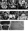
T2-weighted MR imaging of the brain demonstrated replacement of the entire clival bone marrow and cavernous sinus by heterogeneous low signal intensity and small areas of high signal intensity, and there was evidence of sphenoid sinusitis (Figs. 1A-C). The prevertebral muscles showed mixed high and intermediate signal intensity at T2-weighted axial imaging (Fig. 1B), and the pituitary gland was normal in size and shape, with an intact cortical signal void of sellar floor. T1-weighted imaging of the lesion demonstrated mixed intermediate and low signal intensity (Fig. 1D), and at gadolinium-enhanced T1-weighted imaging, intense enhancement was observed (Fig. 1E). The bilateral cavernous sinuses were also involved by the lesion, with diffuse narrowing of the cavernous portion of the right internal carotid artery (Figs. 1A, F). At that time, differential diagnoses included metastatic carcinoma, lymphoma, and an indolent aggressive infectious disease such as fungal infection.
Transsphenoidal clival biopsy was performed, but there was no evidence of fungal infection in the nasal cavity or sphenoid sinus. Biopsy specimens from the clivus revealed only chronic inflammatory cells with thick fibrosis. Immunohistochemistry was positive for vimentin but negative for cytokeratin, CEA, and CD 34. After clival biopsy, a CT examination was performed : the images obtained depicted multifocal permeative bone destruction in the clivus (Fig. 1G) and a bulky retropharyngeal mass of soft tissue density.
A biopsy of the retropharyngeal mass was subsequently perfomed, but the mucosal lining of the nasopharynx revealed no abnormality. Biopsy specimens from the retropharyngeal soft tissue mass-like lesion showed bundles of fibroblasts admixed with inflammatory cells composed of lymphocytes and plasma cells (Fig. 1H). A high-power field view showed no evidence of mitosis or cellular atypism.
The patient was treated with high-dose corticosteroid and discharged after showing clinical improvement.
DISCUSSION
Inflammatory pseudotumor is a chronic inflammatory tumefaction of unknown origin. Because the term 'inflammatory pseudotumor' is nonspecific and lesions have a variety of histologic presentations, several alternative names have been used to refer to them : inflammatory myofibroblastic tumor, plasma cell granuloma or pseudotumor, xanthomatous pseudotumor, pseudosarcomatous myofibroblastic proliferation, inflammatory myofibrohistiocytic proliferation, and myofibroblastoma (1). Although early descriptions of lesions classified as inflammatory pseudotumors focused on their occurrence in the lung, the lesions occur in diverse extrapulmonary locations.
In the head and neck, inflammatory pseudotumor most commonly involves the orbit. According to the pathology literature, a diagnosis of inflammatory pseudotumor of the head and neck has been applied rather indiscriminately. Histopathologically, two characteristics of the lesions have been described. Firstly, the tumor was found to consist of a mixed proliferation of vimentin-positive fibroblasts and smooth muscle actin-positive myofibroblasts, with the former predominating. The cells were arranged in fascicles or sheets, or occasionally in whorls, and mitoses were absent. Secondly, inflammatory cells were present. These lesions probably have a neoplastic origin, and authors have preferred to use the term 'inflammatory myofibroblastic tumor' or 'inflammatory fibrosarcoma' (1).
Reports have stated that extraorbital inflammatory pseudotumors of the head and neck involve the maxillary sinuses (2, 3), infratemporal fossa (4, 5), nasopharynx (including the parapharyngeal space) (4-6), pterygopalatine fossa (6, 7), and the major salivary gland (6). Involvement of the clivus or cavernous sinus by fibrosing inflammatory pseudotumors centered in the nasopharynx, orbit, or masticator space has been described in a previous article (4). To our knowledge, however, a case in which an expansile and infiltrating lesion mainly involved the clivus has not previously been reported.
According to previous reports (2-7), it is very difficult, both clinically and radiologically, to decide whether a lesion involving an infiltrating soft tissue mass, with bony destruction, is a pseudotumor or a malignant neoplasm. In our case, for example, the tumor was at first thought to be a malignant neoplasm such as a lymphoma or metastatic carcinoma, or an aggressive infectious process. There were no signs or symptoms of infection such as fever, and it was unlikely that the heterogeneous signal observed at T2-weighted imaging indicated malignant lymphoma. The coincidence of lesions of differing signal intensity involving the clivus and prevertebral muscle was thought to be unusual for metastatic carcinoma. Without a biopsy, however, differentiation between metastatic carcinoma, lymphoma, and chronic fungal disease was impossible. Multiple biopsies of the clivus and retropharyngeal soft tissue showed only chronic inflammatory cells with thick fibrosis but no mitoses, and the mucosal lining of the nasopharynx was free of both malignancy and infection. Although pathologists eschew the term 'inflammatory pseudotumor' this was our diagnosis on the basis of these findings, and high-dose corticosteroid therapy was indicated.
The administration of high-dose corticosteroid is the primary treatment of choice for inflammatory pseudotumor, and response to medical therapy is thought to roughly parallel the acuity of the inflammatory process. Acute lesions typically respond to high doses of corticosteroid, but chronic lesions, which tend to have more fibrosis, generally do not respond to medical therapy (8). In their series, however, Han et al. (4) found no relationship between the duration of signs and symptoms, and degree of fibrosis. We consider that the findings of MR imaging are a good predictor of therapeutic response. It is postulated that in lesions which show high signal intensity at T2-weighted imaging and strong enhancement, there is a relative abundance of free water and mobile protons. If the response to steroid is poor, complete surgical resection is advocated; when such resection is not possible, local radiation therapy has been shown to be effective in some cases (3, 8).
Where a soft tissue mass involving the clivus shows heterogeneous low signal intensity at T2-weighted imaging, has an infiltrative appearance in the presence of an apparently normal pituitary gland, and there is no known underlying malignancy, inflammatory myofibroblastic tumor, in other words 'inflammatory pseudotumor', should be included in the differential diagnosis.
 XML Download