

 PDF
PDF ePub
ePub Citation
 Print
Print


Since the late 1950s, when A-mode US was first used, ultrasound (US) has been used as a useful diagnostic tool in the evaluation of various ocular pathologies (1, 2). It is especially helpful in patients with vitreous opacity, in which direct visualization of the posterior pole of the eye is impossible (3). The diagnostic criteria of B-mode US for the differentiation of retinal detachment (RD) from vitreous membrane (VM) produced by vitreous hemorrhage are well established (2, 4), but in cases with atypical findings, it is difficult to differentiate the two pathologies using gray-scale US only.
In addition to gray-scale US, Doppler US has been used for periorbital vessels since the 1960s, and color Doppler US has played an important role in the evaluation of orbital and periorbital vascular pathologies (1, 5). In 1991, Wong et al. used color Doppler US to differentiate RD from VM, reporting 100 % sensitivity in the detection of color signals in seven cases of RD (3). Prior to this study, however, we encountered cases in which no color signal was apparent in the detached retina, and were unable to locate any report which described the use of contrast enhancement to distinguish RD from VM. We therefore evaluated the clinical utility of contrast-enhanced color Doppler US for this, comparing it with various conventional US modalities, and analysed the enhancement patterns observed in cases in which there was an enhancement effect.
MATERIALS AND METHODS
Among 40 patients who underwent contrast-enhanced color Doppler US due to vitreous opacity during a recent two-year period, 32 patients [M:F=14:18; age:15-79 (mean, 50.9) years] (32 eyes) were included in the study. There were 14 cases of RD and 18 of VM, and among the former, seven were combined with VM and one with choroid hemangioma. The diagnoses were confirmed by surgery in 28 patients, and by clinical follow-up in two with RD and two with VM.
US examinations were performed by one practitioner using a 10-5 MHz broadband linear array transducer (HDI 5000; ATL, Bothell, Wash). Patients were asked to lie in the supine position with closed eyes and to gaze straight ahead while restricting their ocular movements. After covering the face with a folded towel, a 1-2-cm-thick layer of gel was applied to the eyelid of the examination site to avoid air gaps between the flat surface of the transducer and the curved margin of the lid, and to prevent direct contact between this and the probe. The examiner rested his elbow on the pillow beside the patient's body and his wrist on the folded towel, and placed the probe on the gel with as little pressure as possible. For scanning, the gray-scale mode was used, and the sequence followed was 'horizontal, vertical, oblique', followed by eyeball movement. Color Doppler US was then performed with the color box set at a size which covered the entire area of the vitreous cavity and posterior pole of the eyeball, and at the following settings: color gain from 68% to 82%, wall filter at 'low' or 'medium' level, PRF at 700-1000 Hz, MI (mechanical index) at 0.6-0.8, and flow option in the 'MedV' (medium velocity) or 'HRes' (high resolution) mode. Power and color Doppler US were performed at the same settings.
For contrast-enhanced color Doppler US, 2.5 g Levovist (Schering, Berlin) was injected by hand for 30 seconds at a concentration of 300 mg/mL via an antecubital vein using a 20-22-gauge peripheral intravenous over-needle sheath. Color Doppler examination was begun at the start of contrast injection, and except for color gain, which was controlled within a narrow range according to the degree of 'blooming' artefacts produced by the contrast agent, the color settings were fixed at pre-contrast levels. The process was recorded by a super VHS video tape recorder, and some important images were stored on a hard disc using the cineloop mode.
Gray-scale images were interpreted by two radiologists, who reached a consensus. During all Doppler studies, the diagnostic criterion for RD and VM was whether or not color signals were visualized in the membranous structure(s).
RESULTS
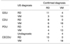
For each US modality, the results of diagnosis are detailed in Table 1.
Gray-scale US correctly diagnosed RD in 11 of 14 cases and VM in 14 of 18; the accuracy with which RD and VM were differentiated was thus 78% (25/32). Pre-contrast color Doppler US depicted color signals in eight eyes with RD and in no eyes with VM, showing a sensitivity of 57% (8/14) in detecting color signals in RD, a positive predictive value of 100% (8/8), and an accuracy of 81% (26/32) in differentiating RD from VM. At power Doppler US, six eyes with RD and one with VM showed color signals, and in eight eyes (RD in 4, VM in 4) diagnosis was unsuccessful due to numerous motion artefacts. Sensitivity was thus 43% (6/14), positive predictive value was 86% (6/7), and accuracy was 59% (19/32). Contrast-enhanced color Doppler US showed color signals in 13 eyes with RD and in no eyes with VM; sensitivity was thus 93% (13/14), with a positive predictive value of 100% (13/13) and an accuracy of 97% (31/32).
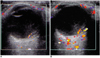
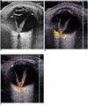
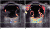
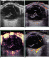
Enhancement patterns were as follows: signal accentuation (n=3), in which only accentuation of the pre-existing color signal seen at pre-contrast color Doppler US was observed (Fig. 1); signal extension (n=2), in which the length of the signal was increased, with or without accentuation (Fig. 2); signal addition (n=3), in which not only the pre-existing signal was observed, but also an additional signal in other part(s) of the membranous structure, with or without signal extension (Fig. 3); and new signal visualization (n=5), which depicted color signals in cases in which a color signal was not seen at pre-contrast color Doppler US (Fig. 4). Enhancement began 12-21 seconds after the start of injection, and its effect lasted for 2-5 minutes.
DISCUSSION
The retina is a structure which is attached to the optic disc posteriorly and ora serrata anteriorly. If the retina is for any reason detached, it is seen at US as a curvilinear structure in the vitreous cavity, attached to the optic disc posteriorly and ora serrata anteriorly, with moderately strong echogenicity and relatively even thickness. In cases of complete RD, it has a 'funnel' or 'morning glory' appearance, and in the chronic state, movement of the detached retina is restricted by movement of the eye. In contrast to RD, VM is not usually attached to the optic disc or ora serrata, and there is weaker echogenicity, less even thickness and greater mobility than in RD (2-4).
Gray-scale US is able to differentiate between the two pathologies using the diagnostic criteria described above. They are not, however, easy to differentiate if RD is partial, shallow, or peripheral, if VM is attached in the vicinity of the optic disc or ora serrata, or if VM is accompanied by RD. Indeed, in our study, the rate of misdiagnosis at grays-cale US was 22% (7/32), which is higher than the rate of 17% (4/24) recorded by Wong et al (3).
In cases with atypical findings at gray-scale US, color Doppler US can play an additional, more reliable role in diagnosis, through visualization of color signals in the detached retina. With regard to retinal blood supply, the inner two-thirds of the retina is nourished by the central retinal artery and the outer one-third by the posterior ciliary arteries. The central retinal artery arises from the ophthalmic artery and enters the optic nerve about 1 cm behind the eyeball. After entering the optic nerve, it is located at the center of the optic disc, where it divides into an upper and lower branch. Each of these then divides into a nasal and a temporal branch, forming superior/inferior nasal and temporal arterioles (1, 3). Color Doppler US can depict the central retinal artery and at least some part of its ramifications, so visualization of a color signal along the detached retina can be expected, with no color signal in VM patients without diabetes.
However, because it depends on factors which include flow velocity, vessel size, the insonation angle of a sound beam, the depth of a lesion, scanner sensitivity, and operator control, color Doppler US is limited in its ability to demonstrate flow in a vascular structure. Accordingly, not all RD cases should be expected to show a color signal in the detached retina. In our series, pre-contrast color Doppler US showed a color signal in only 57% (8/14) of RD cases, though Wong et al. reported 100% (7/7) sensitivity (3). The reasons for the lower detection rate in our series, in spite of the use of a higher resolution scanner, may be the different number of cases and the different conditions of the patients (one-half of all RD cases were combined with VM and six patients were in a post-traumatic and/or postoperative state at the time of examination).
In addition to color Doppler US, power Doppler US is currently widely used in the evaluation of vascular structures, which are displayed by the integrated power of the Doppler signal instead of the mean Doppler frequency shift. Because of increased gain (10 to 15 dB), substantial cutoff of background noise, angle independence and more available dynamic range, power Doppler US is more sensitive than color Doppler US in the depiction of the blood flow signals (6, 7).
However, when power Doppler US was used in this study, numerous artefacts were produced both by the anterior compartment of the globe, including the eyelid, and by the echogenic reflectors in the vitreous cavity (Fig. 4C). The artefacts obscured true signals in the membranous structures, and it was difficult to differentiate them from true signals. Consequently, the use of power Doppler US led to a pseudo-positive finding in one patient with VM and was undiagnostic in eight, and its diagnostic accuracy, at 59%, was thus the lowest among the four US modalities employed in this study. This high number of artefacts was thought to be caused by the high susceptibility of power Doppler US to the inevitable motion of the examiner's hand and a patient's involuntary eyeball movement during examination.
Among other investigations of the application of power Doppler US to the orbit, a study by Giovagnorio and Quaranta reported its superiority over color Doppler US in the evaluation of normal orbital vessels (7). Because the normal vessels they treated are located along the posterior margin of, or beyond, the eyeball, where they are far from the effect of the artefacts produced by its anterior compartment, we agree with their findings, despite our results.
To increase the detectability of Doppler signals, several kinds of the US contrast agents have been developed, and their use in various organs is under investigation.
Levovist is a kind of free-bubble agent consisting of air, palmitic acid-which forms a thin film surrounding the air bubbles, allowing them to remain stable in the body for several minutes-and galactose, which acts as a carrier for the microbubbles. In general, intravascular injection of a US contrast agent increases the number of scatters in the blood, thus increasing the amplitude of the back-scattered signal by 20-30 dB (8). Accordingly, contrast-enhanced Doppler US can be expected to promote the detection of real, but weak, previously undetected high-velocity arterial Doppler signals (9).
With regard to the use of a contrast agent in the evaluation of intrabulbar pathologies, Lemke et al (10). reported that in patients with uveal melanomas, the detection rate of tumor-related vessels by color Doppler US after contrast injection increased only slightly, from 36 of 40 to 38 of 40.
However, when we used contrast media in the detection of a normal vessel in a detached retina, the effect was remarkable.
In our series, five cases of RD in which no color signal was observed at pre-contrast color Doppler US demonstrated new signals in the detached retina at post-contrast color Doppler US, thus significantly increasing the detection rate from 57% (8/14) to 93% (13/14).
In the other eight cases with visible color signals at precontrast color Doppler US, diagnostic confidence increased due to the enhancement effect in a pattern in which the pre-existing color signal was accentuated, extended, or added to.
However, among echogenic materials other than tumors in the vitreous cavity, not only detached retina but also persistent hyperplastic primary vitreous (PHPV) and VM in patients with diabetes can show color signals (3, 11).
PHPV is caused by the failure of normal regression of the components of primary vitreous, including the embryonic hyaloid vascular system. It is associated with the proliferation of embryonic connective tissue forming a mass lesion, and often accompanies retinal detachment due to organizing hemorrhage and retraction (11). Consequently, color signals can be emitted by a persistent hyaloid artery traversing the mid-portion of the mass lesion.
At US, PHPV can be differentiated from RD by its characteristic contour (a triangular-shaped mass), the course of the color signal (extending from the optic disc to the lens), and its occurrence in young children.
In diabetic patients with VM, neovascularity can occur in the membranes due to proliferative diabetic retinopathy. Although the two cases of VM in our series co-occurring with diabetes showed no color signal at either pre- or post-contrast color Doppler US, visualization of a color signal at post-contrast study in patients with diabetes should not be interpreted as RD, while the absence of a visible color signal in both studies could indicate VM.
In conclusion, contrast-enhanced color Doppler US, with an accuracy of 97%, was the most accurate US modality for differentiating RD from VM. In addition, contrast enhancement increased the signal detection rate in RD from 57% (8/14) to 93% (13/14).
 XML Download