

 PDF
PDF ePub
ePub Citation
 Print
Print


The migration of Ascaris lumbricoides into the biliary tree is an uncommon but well-known complication of intestinal ascariasis and often results in biliary colic. The radiologic findings of biliary ascariasis have been described sporadically in the literature (1-3), which has stated that ultrasonography (US) is useful in identifying worms inside the biliary tree (3). We describe two cases of biliary ascariasis undetected by both US and CT but correctly diagnosed after magnetic resonance (MR) cholangiography.
CASE REPORTS
Case 1
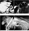
A 44-year-old woman was admitted with a history of epigastric pain which had started ten days prior to admission and had increased in intensity, and associated anorexia, nausea, and fever. Physical examination revealed tenderness in the epigastrium, though liver function tests revealed nothing abnormal and serum bilirubin, amylase and lipase levels, as well as her blood leukocyte count, were within normal limits. US showed no abnormal findings, but contrast-enhanced CT scanning revealed showed dilation of the intrahepatic bile ducts in the left lobe and slightly hyperattenuating lesions within the dilated bile ducts (Fig. 1A). MR cholangiography involving projection imaging and single-shot rapid acquisition using the relaxation enhancement (RARE) technique (repetition time, ∞ msec; effective echo time, 1200 msec; echo spacing, 11.5 msec; echo train length, 240; flip angle, 150°; slab thickness, 70 mm; field of view, 300 mm; number of signals acquired, one; matrix, 240×256; and acquisition time, 6.32 sec) showed a hypointense tubular filling defect in the left intrahepatic bile duct (Fig. 1B). Endoscopic retrograde cholangiography demonstrated a linear filling defect along the left intrahepatic and common bile duct (Fig. 1C).
The worm was endoscopically removed through the ampulla of Vater (Fig. 1D), no additional treatment being undertaken. The patient's clinical symptoms subsequently subsided.
Case 2
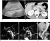
A 41-year-old woman was admitted with a ten-day history of epigastric pain. Physical examination revealed tenderness in the epigastrium, and laboratory tests indicated elevated serum amylase (2504 U/L) and lipase (2535 U/L) levels. The results of liver function tests were normal, and her serum bilirubin level and blood leukocyte count were within normal limits. US revealed slight enlargement of the pancreas and mild dilation of the bile ducts (Fig. 2A). Other than a slightly enlarged pancreas, contrast-enhanced CT scanning demonstrated no abnormality (Fig. 2B). An MR cholangiogram obtained by projection imaging using the single-shot RARE technique showed a hypointense tubular filling defect in the common bile duct (Fig. 2C). Successive MR cholangiograms obtained in the same area every 30 seconds for 15 minutes demonstrated the movement of a worm within the bile duct (Fig. 2D). Endoscopic retrograde cholangiography showed a linear filling defect in the common bile duct (Fig. 2E), and the worm was removed endoscopically. In addition, albendazole was administered orally and the patient's clinical symptoms subsequently improved.
DISCUSSION
Ascaris lumbricoides is one of the most common worldwide causes of human helminthic infestation (1). An adult worm is typically 15-50 cm long and 3-6 mm thick, and the clinical presentation usually involves the passage of worms through the rectum, or vomiting, and small bowel obstruction by a bolus of worms. The migration of a worm through the papilla of Vater into the biliary tree is an uncommon complication of ascariasis and leads to biliary colic, recurrent pyogenic cholangitis, pancreatitis, hepatic abscesses and septicemia.
If biliary ascariasis is suspected, US is the imaging modality of choice. The US findings of biliary ascariasis have been previously described: tubular, echogenic, non-shadowing structures, sometimes with a thin, longitudinal, central sonolucent line (2-4, 6, 8). Unless the operator carefully examines the biliary tract, however, the diagnosis of biliary ascariasis is difficult and in this respect, endoscopic retrograde cholangiography is a good but relatively invasive tool (4, 8-10). Several reports have described the MR imaging findings of biliary ascariasis (4, 6-7): on T1-weighted images, worms are seen as linear, slightly hyperintense tubular structures with a central hypointense area (7) and where there is massive biliary ascariasis, MR cholangiography shows intraductal worms as linear hypointense filling defects in the biliary tract (6).
In our two cases, CT and US failed to diagnose biliary ascariasis, though subsequent MR cholangiography clearly demonstrated a linear filling defect within the bile duct that led to the correct diagnosis and proper treatment. State-of-the-art MR cholangiography using the single-shot RARE technique provided excellent images, comparable to those obtained by endoscopic retrograde cholangiography. In one case, MR cholangiography repeated at 30-second intervals demonstrated movement of a live worm within the bile duct.
In conclusion, MR cholangiography using the single-shot RARE technique is a valuable non-invasive tool that clearly demonstrates biliary ascariasis.
 XML Download