

 PDF
PDF ePub
ePub Citation
 Print
Print


Gangliocytomas of the central nervous system are very rare, and tend to occur in children and adults under 30 years of age (1). They represent one type of ganglion cell tumors and are composed of mature ganglion cells. This pathologic feature distinguishes gangliocytomas from gangliog1iomas, which contain both ganglion cells and g1ia1 cells (2). Supratentorial gangliocytomas have rarely been reported and have not been clearly documented (3, 4). We describe two cases of supratentorial gangliocytoma in which unusual MR findings mimicking extra-axial tumors such as meningiomas were observed.
CASE REPORTS
Case 1
A previously healthy 22-year-old woman initially presented with right hemiparesis and episodes of right-sided sensory abnormalities, both of which had begun one year earlier. She also had a one-year history of intermittent loss of consciousness unrelated to these symptoms. General physical examination and the neurological findings were unremarkable. Routine electroencephalography (EEG) demonstrated diffuse cerebral dysfunction over the left hemisphere, but no epileptic or epileptiform discharge was noted.
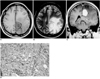
MR studies of the brain using a 1.5-T system revealed a well demarcated broad-based mass, 2 cm in size, on the falx cerebri and prominent peritumoral edema. Realative to gray matter, the mass was hypointense on T1-weighted MR images (Fig. 1A) and hyperintense on T2-weighted images (Fig. 1B). Gadolinium-enhanced T1-weighted images demonstrated strong homogeneous enhancement (Fig. 1C). Along its lateral margin, the tumor's cystic structure was isointense relative to cerebrospinal fluid, and it was thus thought to be a peritumoral cyst (Figs. 1A, B).
Surgery revealed that the tumor was located in the brain parenchyma, adhered to the falx cerebri, and had an extra-axial component. Its gross appearance was slightly lobulated and it was whitish gray in color. There was no necrosis or calcification within the mass, and microscopic examination showed that it was composed of relatively mature ganglion cells and abundant collagen-rich stroma in which spindle stromal cells and lymphocytes were observed (Fig. 1D). Both the cytoplasm of the ganglion cells and stromal cytoplasmic processes stained positively with antibody for synaptophysin, though glial fibrillary acidic protein (GFAP) staining failed to reveal a glial component. Finally, gangliocytoma was diagnosed.
Case 2
A previously healthy 59-year-old woman presented with stubborn headache and dizziness which had begun six months earlier. She also complained of intermittent facial palsy and had a seven-year history of hypertension. The results of neurological examination were perfectly normal, and routine EEG showed no epileptic or epileptiform discharge.
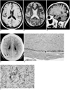
MR images of the brain demonstrated a small dural-based mass, and adjacent to it, in the right parieto-occipital region, was a small amount of subdural hygroma. Relative to gray matter, the mass was hypointense on T1-weighted images (Fig. 2A) and hyperintense on T2-weighted images (Fig. 2B). On gadolinium-enhanced T1-weighted images, homogeneous enhancement was observed (Fig. 2C). The tumor appeared to have both intra- and extra-axial components; tumor-cell infiltration of brain parenchyma was thought to arise from the intra-axial portion (Fig. 2B). Gadolinium-enhanced T1-weighted sagittal images showed that the major portion of the tumor was located intra-axially (Fig. 2C), and CT scanning demonstrated calcification within the tumor (Fig. 2D). The preferred preoperative radiologic diagnosis was an extra-axial mass such as atypical meningioma.
Surgery revealed that the tumor was located in the cortical area of the brain parenchyma, showed partly exophytic growth, and adhered to the adjacent leptomeninges. The clinical symptoms resolved postoperatively. Grossly, the tumor was well demarcated. Its cut surface was soft and whitish, and it contained yellowish friable granular material. Microscopic examination showed that it was composed of ganglion cells, and there was marked desmoplastic reaction and lymphocytic infiltration. The ganglion cells varied considerably in size and shape, but a glial component was not clearly demonstrated. Numerous eosinophilic granular bodies were also observed. Microscopic examination (low-power field) showed that the mass was attached to the dura mater (Fig. 2E).
Immunohistochemistry demonstrated positive staining of the cytoplasm and process of some tumor cells with synaptophysin, but negative staining with GFAP.
DISCUSSION
Ganglion cell tumors include gangliogliomas, gangliocytomas, dysplastic cerebellar gangliocytomas (Lhermitte-Duclos disease) and desmoplastic infantile gangliogliomas (2). Among these, gangliogliomas and gangliocytomas are not always distinct. A tumor is designated 'ganglioglioma' if it has a neoplastic glial component, while one in which only abnormal neurons are present is a gangliocytoma. Considering the group of differentiated ganglion cell tumors of the CNS as a whole, it is apparent that in the glial component there are varying degrees of neoplastic evolution, and this variability makes clear-cut separation of gangliocytomas and gangliogliomas difficult. In the two cases described in this paper, the tumors were composed mainly of ganglion cells, while immunohistochemical staining for GFAP failed to unequivocally identify a glial component. In both cases, 'gangliocytoma' is thus an appropriate designation.
Gangliocytomas are extremely rare, with a reported frequency of only 0.1% (5). They are almost always benign, so preoperative study of their radiological features is important (2). Supratentorial gangliocytomas have on occasion been cited as the cause of epileptic foci (3, 6), though discussion of their specific clinicoradiological features has been limited to individual case reports (3, 4, 7).
Previous reports have failed to document clearly the imaging findings of supratentorial gangliocytoma. According to reports by Sherazi and Peretti-Viton et al. (4, 7), MR imaging of gangliocytomas revealed masses of low signal intensity on T1-weighted images and high signal intensity on T2-weighted images, while gadolinium-enhanced scanning frequently demonstrated enhancement. In terms of MR signal intensity and degree of contrast enhancement, our two cases were similar to those of Sherazi and Peretti-Viton et al. Altman (3), however, reported that MR imaging of supratentorial gangliocytomas demonstrated intra-axial tumors of mixed signal intensity on T1-weighted and proton-density images, low signal intensity on T2-weighted images, and little mass effect in three cases involving children with intractable seizure. Altman suggested that the low signal intensity seen on T2-weighted images might be the result of large nuclei and prominent nucleoli with long-chain nucleic acids. The high signal intensity seen on T2-weighted images, as in our two cases and two previous reports (4, 7), might be attributed in part to the abundant cytoplasm of the ganglion cells. Peretti-Viton et al. reported that CT scanning of these tumors often revealed calcification and cysts. In our second case report, calcification was observed.
In both our cases, tumor location was also quite interesting. Both tumors were located peripherally, with some exophytic components. In the first case, a broad-based mass was attached to the falx cerebri and had a dural tail, mimicking falx meningioma. In the second case, the tumor showed partly exophytic growth, mimicking an extra-axial tumor, and adjacent to it there was a small amount of subdural hygroma. According to Altman and Peretti-Viton et al. (3, 7), supratentorial gangliocytomas occurred in cortical and subcortical locations. In the case described by Itoh et al. (8), the gangliocytoma was also cortically located. In previous reports (3, 7, 8) and in our cases, tumors were peripherally located. This predominantly cortical location might be because the tumor originated from ganglion cells in the cortex. In previous reports, however, MR findings suggesting an extra-axial or exophytic component such as a dural tail or subdural hygroma, as in our cases, were not mentioned, and the tumors seen on CT or MR images were evidently considered to be intra-axial. The dural tail of the tumor in our first case might have resulted from fibrous adhesion to the falx. In both our cases, microscopic examination revealed that tumor cells were diffusely positive for Masson's trichrone staining and there was a marked desmoplastic reaction, characteristics which might result in fibrous adhesion to the falx or meninges. The subdural hygroma adjacent to the tumor, as seen in our second case, might be secondary to the exophytic component of the tumor and the fibrous adhesion to the leptomeninges. We believe that the presence of these extra-axial components and fibrous adhesion to the falx or leptomeninges could be important imaging findings of supratentorial gangliocytoma.
The differential diagnoses of supratentorial gangliocytoma include ganglioglioma or meningioma, but a rigid separation between gangliocytoma and ganglioglioma on either histological or clinical grounds is difficult. As already described (4, 7), the MR and CT findings of gangliocytomas showed significant overlap with those of gangliogliomas, and differentiation between them on the basis of imaging findings is thus difficult or impossible. Differentiation from meningiomas can sometimes be problematic, as in our cases. Since meningiomas usually show iso or low signal intensity on T2-weighted images, high signal intensity in these circumstances can provide a means of differentiation.
In summary, supratentorial gangliocytoma, a very rare benign neuronal tumor, can manifest as a cortical or subcortical lesion which shows high signal intensity on T2-weighted MR images and homogeneous enhancement on contrast-enhanced images, and calcification may be observed. It can, however, as we report here, mimick an extra-axial tumor such as meningioma.
 XML Download