

 PDF
PDF ePub
ePub Citation
 Print
Print


INTRODUCTION
Lateral patellar dislocation (LPD) is a common injury that typically occurs in young, active patients as a result of a variety of activities and accounts for approximately 2–3% of all knee injuries (1). Previous studies have shown that anatomic factors such as patella alta, increased tibial tubercle-trochlear groove (TT-TG) distance, rotational deformity and trochlear dysplasia are all associated with an increased risk of a recurrent LPD (2345678910). Although injury to the medial patellofemoral ligament (MPFL) may reduce passive stability and predict subsequent instability with non-operative treatment (111213141516), the correlation between injury patterns of MPFL (injury severity and location) in acute first-time LPD and incidence of a recurrent (second-time) LPD has not been fully and clearly clarified (91516). Thus, the purpose of this study was to use qualitative and quantitative methods to clarify the correlation between the injury patterns of MPFL in an acute first-time LPD and the incidence of second-time LPD at 5-year follow-up.
Go to : 
MATERIALS AND METHODS
Patients
This study was approved by our Institutional Ethics Committee, and informed consent was obtained from all patients. Patients were eligible for inclusion if they had suffered an acute, first-time LPD regardless of the injury mechanism. The inclusion criteria were as follows (1718192021):
1) the presence of a locked acute dislocation or history of reduced dislocation within 15 days of the injury; 2) typical clinical findings including haemarthrosis, medial parapatellar structures, femoral epicondyle that is painful on palpation, and apprehension sign (except in locked dislocation) are present; and 3) a bone contusion involving both the lateral femoral condyle and medial patella as demonstrated on the MRI image.
The exclusion criteria included previous surgery on the injured knee, conditions associated with serious neuromuscular or congenital diseases, and a knee joint with a multiple-ligament injury. When no distinct fibers or remnants were identified at the expected MPFL course on the magnetic resonance (MR) image and there was no appreciable surrounding edema, the ligament was designated as absent and the case was excluded from the study. In addition, the purpose of this study was to investigate long-term nonoperative healing of the injured MPFL; thus, we excluded patients who had undergone patellofemoral surgery (MPFL repair or MPFL reconstruction) after the first-time LPD. However, the patients who only had undergone an arthroscopic operation to remove the osteochondral loose bodies were included in this study.
The selected patients were voluntarily participating in a five-year follow-up study at our hospital. All patients underwent a clinical examination in our orthopedics department six months after the first-time LPD, and then were interviewed by telephone every three months. The follow-up was performed by an orthopedic surgeon in our study group. The final interview was conducted at 60 months after the first-time LPD. During the follow-up, patients who had experienced knee injuries were examined and cured in our hospital. If the LPD was a relapse (per the criteria mentioned above), the time interval between the two LPDs was recorded. The injury patterns of MPFL in the intraoperative findings were compared with the preoperative MRI examination in patients who were surgically treated (MPFL suture or reconstruction) in the follow-up because of a recurrent LPD. Moreover, during the follow-up, patients were excluded from the study because of other injuries or diseases that occurred between the two LPDs or occurred in the non-recurrent LPD subgroup that prevented the knee joint from returning to its optimal status.
Using the criteria mentioned above, the study enrolled 147 knees of 147 consecutive patients with a mean age of 20 years (range, 8–42 years; 80 women [mean age, 19 years; range, 8–41 years] and 67 men [mean age, 22 years; range, 9–42 years]).
MRI Technique
The MRI was performed on a 1.5-tesla system (Magnetom Symphony Syngo MR A30; Siemens, Erlangen, Germany), and all patients underwent imaging with their knees positioned in full extension. The following sequences were performed: a transverse fat-saturated proton-density weighted fast spin-echo imaging sequence (repetition time/echo time, 4500 ms/33 ms; flip angle, 150°; field of view [FOV], 160 × 160 mm; section thickness, 3.0 mm) was followed by a coronal fat-saturated proton density (2000/15; FOV, 150 mm; matrix, 320 × 224 pixels; slice thickness, 3.0 mm; skip, 0.3 mm), a sagittal proton density (1950–2766/14; FOV, 140–150 mm; matrix range, 320–384 × 192–224 pixels; slice thickness, 3.0 mm; skip, 0.3 mm), a sagittal fat-saturated proton density (2650–4366/13–16; FOV, 140–150 mm; matrix range, 256–384 × 224–256; slice thickness, 3.0 mm; skip, 0.3 mm), and a sagittal T1-weighted sequence (450–600/10–20; FOV range, 140–150 mm; matrix range, 320–384 × 224; slice thickness, 3.0 mm; skip, 0.3 mm).
MRI Evaluation
The MR images were analyzed independently by two radiologists who had 13 years and 16 years of clinical experience in musculoskeletal radiology, respectively, and were both unaware of the results of the previous imaging interpretations. The conclusions of each radiologist were initially recorded. In the event of disagreement, the images were then reviewed to reach consensus.
Using the diagnostic classification criteria from studies of Zhang et al. and Balcarek et al. (1718192021222324), the degrees of MPFL injury were divided into partial and complete tears. The manifestations of a partial MPFL tear were defined as thickening and irregularity of the contour, including discontinuity of normal fibers, and intraligamentous or extensive periligamentous edema. The manifestations of a complete MPFL tear were defined as completely discontinuous or apparently absent fibers in the expected region of the MPFL with extensive surrounding edema. Partial or complete disruption of the MPFL was evaluated at 3 locations: the patellar insertion (PAT), the mid-substance (MID), and the femoral attachment (FEM). An evaluation was also performed on avulsion-type fractures at the PAT or FEM. Simultaneous injury at more than one location of the MPFL was classified as a combined injury (COM) (17181920212223).
Trochlear dysplasia, which is dysplasia of the femoral trochlea, was categorized as normal, low-grade trochlear dysplasia or high-grade trochlear dysplasia for further analysis (25). The patellar height was evaluated on the sagittal MR images according to the index of Insall and Salvati (i.e., the patellar tendon length to the longest sagittal dimension of the patella). Patella alta was considered with ratios of 1.3 or greater (252627). The TT-TG distance was assessed in conformity with Schoettle et al. (28). A TT-TG distance greater than 20 mm was classified as abnormal (41027).
Statistical Analysis
Data analyses were performed using SPSS version 17.0 software (SPSS Inc., Chicago, IL, USA). The mean value, standard deviation, and range are presented. Independent samples t tests, chi-square tests and Kruskal-Wallis tests were used to compare the variables in the study subgroups (as described later). A p value of less than 0.05 was considered statistically significant.
Go to :
RESULTS
Injury Patterns of MPFL in Acute First-Time LPD
An injury to the MPFL was found in 142 patients (96.6%) after an acute first-time LPD, including 62 cases with a partial tear (42.2%) and 80 cases with a complete tear (54.4%). For the remaining 5 patients (3.4%), no obvious MPFL injury was identified. Of the 80 cases with a complete MPFL tear, 11 of them included an avulsion-type fracture at the patellar insertion and 3 of them included an avulsion-type fracture at the femoral attachment.
Based on the injury patterns of the MPFL (injury location and severity) of an acute first-time LPD, the study group was divided into 2 subgroups for further analyses: injury location (FEM, PAT, or COM) and injury severity (partial or complete tear). Patients with an isolated injury at the MID of the MPFL (4 patients) were excluded from the statistical analyses of the injury location subgroup because of the small sample size. The subgroups were considered parametric in relation to age and sex; there were no statistically significant differences between the groups. The results of the correlation analyses between the injury patterns of MPFL in an acute first-time LPD and anatomic variants of the patellofemoral joint are shown in Tables 1 and 2.
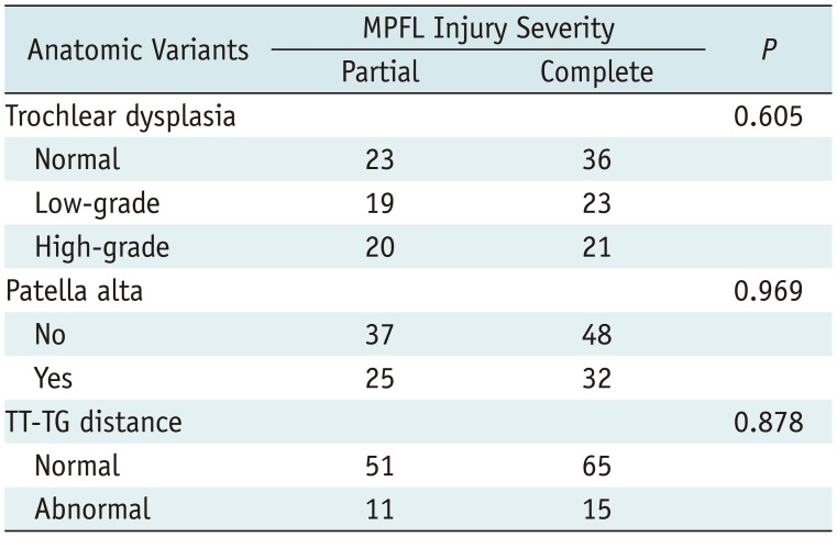
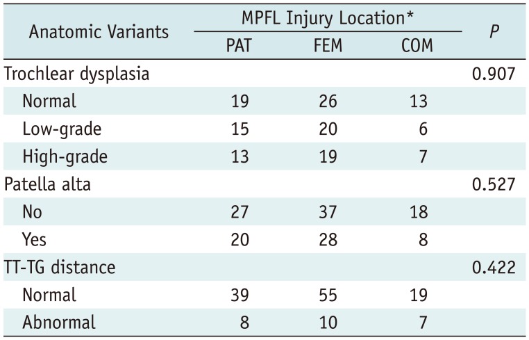
Table 1
Statistical Analysis between Subgroups of MPFL Injury Severity in First-Time LPD and Anatomical Variants
![]()
Table 2
Statistical Analysis between Subgroups of MPFL Injury Location in First-Time LPD and Anatomical Variants
*Patients with isolated lesion at mid-substance (four patients) were excluded from statistical analysis. Five patients without obvious MPFL injury were excluded from statistical analysis. COM = injuries occurred simultaneously in more than one location, FEM = isolated femoral attachment, PAT = isolated patellar insertion
![]()
Second-Time LPD
In the ensuing 5-year follow-up, the incidence rate of second-time LPD was 31.3% (46 of 147). The time interval between the two LPDs ranged from 7 to 56 months. The injury locations of MPFL in the second-time LPDs were exactly the same as those of the first-time LPD (100%).
Diagnostic Performance of Preoperative MRI
Of the 24 patients who underwent surgery (MPFL suture or reconstruction) after a second-time LPD in our study, 18 had a complete MPFL tear and 6 had a partial MPFL tear. Overall, 28 localizations of MPFL injury were present in 24 patients. In a site-based analysis, the diagnostic accuracy of MRI was 100% for partial MPFL tears and 100% for complete MPFL tears.
Correlation Analyses between Injury Patterns of MPFL in Acute First-Time LPD and the Incidence of Second-Time LPD
The results of the correlation analyses of the injury patterns of MPFL in acute first-time LPD and the incidence of second-time LPD are shown in Tables 3 and 4.
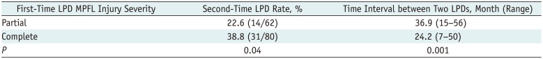
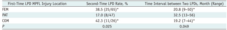
Table 3
Statistical Analysis between Subgroups of MPFL Injury Severity in First-Time LPD and Incidence of Second-Time LPD
| First-Time LPD MPFL Injury Severity | Second-Time LPD Rate, % | Time Interval between Two LPDs, Month (Range) |
|---|---|---|
| Partial | 22.6 (14/62) | 36.9 (15–56) |
| Complete | 38.8 (31/80) | 24.2 (7–50) |
| P | 0.04 | 0.001 |
![]()
Table 4
Statistical Analysis between Subgroups of MPFL Injury Location in First-Time LPD and Incidence of Second-Time LPD
![]()
The incidence rate of second-time LPD in the complete tear subgroup was statistically higher than that of the partial tear subgroup at the 5-year follow-up (38.8% vs. 22.6%, p = 0.04). The mean time interval between two LPDs in the complete tear subgroup was significantly shorter than that of the partial tear subgroup (24.2 months vs. 36.9 months, p = 0.001) (Table 3). The incidence rates of second-time LPD in the FEM and COM subgroups were significantly higher than that of the PAT subgroup (38.5 and 42.3% vs. 17.0%, p = 0.025). In addition, the mean time intervals between two LPDs were significantly shorter in the FEM and COM subgroups than that of the PAT subgroup (20.8 and 19.2 months vs. 32.5 months, p = 0.049) (Table 4). Representative cases are shown in Figures. 1, 2, 3, 4
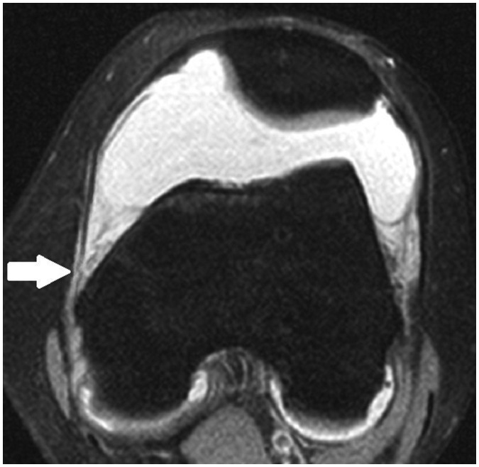
 | Fig. 114-year-old girl was diagnosed with partial MPFL tear at its patellar insertion in acute first-time LPD.After 22 months, she had recurrent LPD. Axial fat-saturated proton density-weighted fast spin-echo imaging sequence shows irregularity with intraligamentous edema of MPFL at its patellar insertion (arrow) in first-time LPD. LPD = lateral patellar dislocation, MPFL = medial patellofemoral ligament
|
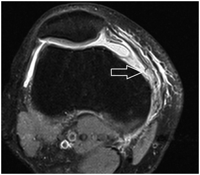
 | Fig. 216-year-old male was diagnosed with partial MPFL tear at its femoral attachment in acute first-time LPD.After 23 months, he had recurrent LPD. Axial fat-saturated proton density-weighted fast spin-echo imaging sequence shows irregularity with intraligamentous and periligamentous edema of MPFL at its femoral attachment (arrow) in first-time LPD.
|
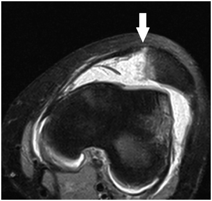
 | Fig. 324-year-old man was diagnosed with complete MPFL tear at its femoral attachment in acute first-time LPD. After 16 months, he had recurrent LPD.Axial fat-saturated proton density-weighted fast spin-echo imaging sequence shows complete discontinuity of MPFL at its femoral attachment with retraction of fibers anteriorly and extensive surrounding edema (open arrow) in first-time LPD.
|
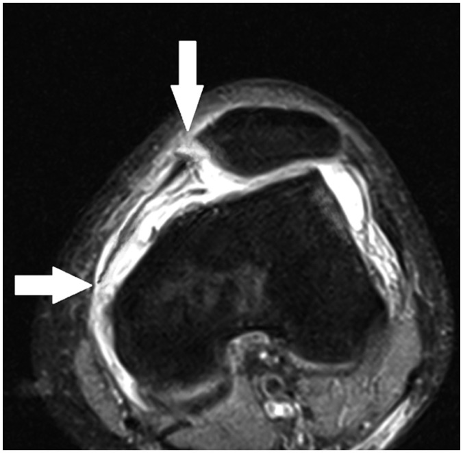 | Fig. 422-year-old woman was diagnosed with combined MPFL injury (complete MPFL tear at its femoral attachment and patellar insertion) in acute first-time LPD.After 7 months, she had recurrent LPD. Axial fat-saturated proton density-weighted fast spin-echo imaging sequence shows complete discontinuity of MPFL at its patellar insertion and femoral attachment with retraction of fibers and extensive surrounding edema (arrows) in first-time LPD. Note also small avulsion fracture from medial margin of patella.
|
Go to :
DISCUSSION
As the most important passive restraint against LPD, the MPFL accounts for 50–60% of the total restraining force against a LPD (29303132). A MPFL injury occurred in up to 78.4–100% of the cases after an acute LPD (17181920212223333435363738). The results of the present study were consistent with those of previous studies that MPFL was more easily injured at the FEM, followed by the PAT (172038). The distribution of the MPFL tears may be related to the anatomy and injury mechanism of the MPFL and the ossification process of the medial patella border and distal femoral epiphysis (1834394041).
Trochlear dysplasia, patella alta and an increased TT-TG distance are primary anatomical variants for a first-time and recurrent LPD (234567891027). Trochlear dysplasia is the main risk factor for a LPD, while patellar alta and abnormal TT-TG distance act as additional risk factors for a LPD (27). Our study results were consistent with the findings of Zhang et al. and Weber-Spickschen et al. that demonstrated there were no correlations between the injury patterns of MPFL and trochlear dysplasia, and patellar height and TT-TG groove distance in a first-time LPD (1942).
The results of the present study showed that recurrent LPD was very common after conservative treatment of the first-time LPD. Moreover, the injury locations of MPFL in the first- and second-time LPDs were exactly the same. This outcome may be an indicator of the poor ability to self-repair and self-heal a torn MPFL. On the other hand, the present study showed that there was a significant difference in the partial and complete MPFL injuries in a acute first-time LPD with respect to the incidence rate of second-time LPD in the ensuing 5-year follow-up. In addition, the mean interval of time between the two LPDs was shorter in the complete MPFL tear subgroup than that of the partial tear subgroup. This finding can be explained by the fact that MPFL is the primary passive restraint against LPD with a poor ability to self-repair and self-heal, as mentioned above. As the severity of the MPFL injury in first-time LPD (complete tear) increases, the stable force to the patellofemoral joint decreases, thereby becoming more predisposed to a recurrent LPD.
In addition, the present study also showed that the isolated femoral-side and combined MPFL tears in an acute first-time LPD also predisposes the patient to a recurrent LPD. This outcome also can be explained by the anatomy of MPFL. The femoral attachment is the weakest part of MPFL. On the contrary, the patellar insertion of MPFL is much thicker than the femoral attachment and is reinforced by the tendon of vastus medialis obliquus muscle, medial retinaculum, and medial patellotibial ligament at the patellar attachment (394041). So compared with the patellar insertion of MPFL, the ability to self-repair and self-heal in the FEM subgroup may be even worse. Therefore, the FEM subgroup is more predisposed to re-dislocation with the same external force. We also surmise that the relevant factors are liable to cause re-dislocation in the COM subgroup. However, the anatomy of patellofemoral joint and the injury mechanisms in a LPD are not yet fully understood. Hence, further studies are needed to confirm these hypotheses.
The appropriate therapy for patients with first-time LPD remains a controversial issue (122637434445464748). According to the optimal available evidence, compared with non-operative treatment, operative treatment of acute LPD may result in a lower recurrence rate of LPD, higher health-related quality of life, and sporting function (434449). Although many anatomic variants of the patellofemoral joint have an impact on the incidence of a recurrent LPD, a MPFL repair or reconstruction remains the primary surgical treatment method (2345678910434449). In fact, MPFL reconstruction has been broadly accepted as the primary surgical treatment for recurrent LPDs with good clinical results (50). Although still in dispute, most studies have suggested that trochlear dysplasia, patella alta and TT-TG distance did not result in a significant difference in the outcomes of an isolated MPFL reconstruction (8515253545556). Therefore, the injury patterns of MPFL in first-time LPD may be another important factor to consider when determining the optimal treatment. When the complete, isolated femoral-side or combined MPFL tears are identified after an acute first-time LPD, surgical treatment, including MPFL repair or reconstruction, may be considered.
This study had some limitations. First, the MPFL injury patterns in acute first-time LPDs were not evaluated surgically but based on the MRI findings only; therefore, it was impossible to verify the diagnostic accuracy of the MRI findings in the present study. Nonetheless, of the 24 patients who underwent surgical treatment after a second-time LPD, the accuracy of the preoperative MRI was 100% in the diagnosis of MPFL injury patterns. In addition, previous studies also have shown that MRI is an accurate method for the diagnosis of the injury patterns of MPFL after an acute LPD (171920225758). Therefore, the present study could be carried out based on the MR images. Second, anatomical factors of the patellofemoral joint are included only as considerations for the subgroups of injury patterns of MPFL in a first-time LPD and not incorporated as independent parameters in this follow-up study. Nonetheless, previous studies have already investigated the correlations between the anatomical factors of the patellofemoral joint and incidence of a recurrent LPD (2345678910). The purpose of our study was just to analyze the correlation between the injury patterns of MPFL in a first-time LPD and incidence of a second-time LPD. Accordingly, the anatomical factors are not taken into consideration in a recurrent LPD. To evaluate the risk factors of a recurrent LPD, further multivariate logistic regressions are required to assess the anatomic factors of patellofemoral joint and injury patterns of MPFL in a first-time LPD. Third, only the incidence of a second-time LPD was analyzed in the present study; the number of LPDs in the 5-year follow-up was not evaluated. It deserves further attention in future studies.
In conclusion, second-time LPD is very common after conservative treatment following an acute first-time LPD. Compared with partial MPFL tears, complete tears predispose recurrent LPD, including a higher incidence rate of second-time LPD and a shorter time interval between dislocations. Compared with the isolated patellar-side MPFL tears, the isolated femoral-side and combined MPFL tears predispose recurrent LPD, including a higher incidence rates of second-time LPD and shorter time intervals between dislocations. Thus, the MPFL injury patterns in an acute first-time LPD may be another factor to consider in treatment selections.
Go to :
 XML Download