

 PDF
PDF ePub
ePub Citation
 Print
Print


INTRODUCTION
Primary neoplastic conditions of the anal canal are uncommon. Although their diagnosis and follow-up are traditionally based on clinical assessment, imaging, particularly MRI, plays an important role in staging and follow-up after treatment. In this article, we discuss and illustrate the current role of MRI in the pre and post-therapeutic management of these patients.
Anatomical and Histological Relevant Notions
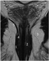
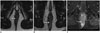
The anal canal extends from the anorectal junction to the anal margin, and is usually 3–6 cm in length. The anal margin is the circular rim of pigmented skin with folds surrounding the anus. The anal canal is composed of an internal sphincter and an external sphincter complex. The internal sphincter is the distal continuation of the inner circular rectal muscular layer, while the external sphincter complex includes the inferior confluence of the levator ani muscle, the puborectalis sling, and the deep, subcutaneous, superficial external sphincter muscles. Both sphincters are separated by the fatty intersphincteric space (Figs. 1, 2).
A significant marker of the anal canal is the dentate line, separating the upper part lined with transitional zone or rectal glandular mucosa, and the lower part (distal third) lined by nonkeratinizing squamous epithelium. Tumors located above the dentate line drain into the perirectal and internal iliac nodes, whereas tumors below the dentate line drain into the inguinal and femoral lymph nodes. The dentate line cannot be seen on MRI, but its position can be estimated by dividing the anal canal into thirds, so that the dentate line lies at the junction of the upper one-third and lower two-thirds. The current World Health Organization distinguishes invasive tumors as epithelial tumors (squamous cell carcinoma, adenocarcinoma, mucinous adenocarcinoma, small cell carcinoma, undifferentiated carcinoma), nonepithelial tumors (leiomyoma, gastrointestinal tumor, myofibroma), carcinoid tumors and melanomas. Virtually all anal canal cancers (AC) are squamous, either keratinizing or non-keratinizing, according to their origin below or above the dentate line, although with similar biological behaviour. As far as tumors near the anorectal junction are concerned, the American Joint Committee on Cancer (AJCC) recommends that tumors with an epicenter more than 2 cm superior to the dentate line should be staged as low rectal carcinomas, whereas the others tumors should be staged as AC (1). In practice, however, the precise point of origin is often uncertain at diagnosis.
Epidemiology of Anal Cancer
Anal canal cancers are uncommon, with an incidence of 1.5:100000 persons per year (2). Their rate has been steadily increasing, due to the rising prevalence of the strongest risk factors, the human immunodeficiency virus (HIV), and human papilloma virus (HPV), mainly HPV-16 subtype (3,4,5). Although the overall incidence of AC remains higher among women, it is much higher in men who practice anoreceptive intercourse (4). AC is also strongly associated with immune suppression in transplant recipients (3). Other risk factors include cervical dysplasia, autoimmune disorders and cigarette smoking (4). Some reports have also shown an increased risk of anal cancer with long-standing, severe perianal fistulizing Crohn's disease (5,6,7). In these cases, AC occurs at a younger age. Diagnosis is often delayed, and at an advanced stage the prognosis is grave.
Clinical Diagnosis and Treatment of Anal Cancer
Diagnosis of these tumors is often delayed as they commonly present with bleeding that is generally attributed to hemorroids. They may also manifest as any combination of a mass, non-healing ulcer, pain, itching, discharge, fecal incontinence and fistula. The diagnosis of anal cancer is made on biopsy-proven histology. More advanced lesions in the distal anal canal may extend to the skin at the anal margin. Rarely, patients present with inguinal lymphadenopathy.
Anal margin tumors can be treated by surgical excision alone, as opposed to anal canal tumors, which have a worse prognosis and are treated with definitive chemoradiotherapy.
A multidisciplinary approach is mandatory. Large prospective validated randomized controlled trials (8,9,10,11,12) support the use of radiotherapy with a dose ranging between 45 and 50 Gy, combined with mitomycin-C and infusional 5-FU chemotherapy as the standard of care for all AC. This leads to complete tumor regression in 80–90% of patients, with locoregional failures of up to 15% (13). Standard radiation fields include the pelvis, anus, perineum, and inguinal nodes. Inguinal nodes receive a booster dose if there is evidence of gross nodal disease (14). The incidence of nodal involvement increases with increasing primary tumor size, and is at least 20% in patients with T3 disease (15). In case of surgical treatment, abdomino-perineal resection is performed for salvage in patients with locally recurrent disease, or for non-responsive patients with persistent tumors (13). Local excision may be performed in case of smaller lesions (< 2 cm in diameter), those involving the anal margin, and well differentiated lesions (13). In these cases, it is important to adequately stage the lesion in order to rule out the presence of positive nodes. Lastly, metastatic disease is considered incurable.
MRI Protocol
Most studies dealing with MRI of AC have been performed at 1.5T (14,15,16). No specific patient preparation is required. Although some teams use antispasmolytic agents to obtain good quality MR images (16,17,18,19), we did not find this a necessity. As for rectal cancer (20), insertion of a probe in the anal canal does not seem to be necessary either. Adequate coverage of the anal margin, inguinal areas and higher nodal stations, i.e., at least the sacral promontory and the area below the aortic bifurcation, should be obtained. Some authors also add T1-weighted MR sequences up to the renal hilum (21). MR protocols include high-resolution T2-weighted sequences along the three planes, with coronal and axial scans planned parallel and perpendicular to the long axis of the anal canal. Slice thickness for imaging the tumor is generally kept under 4 mm (16,17,18,19, 22, 23), with a small field of view.
Most authors (16,17,18,19,20) use additional short-tau inversion recovery sequences. Despite their limited spatial detail, these sequences are helpful for identifying fistula tracts present at the initial staging, or those developing during the treatment. We also use gadolinium-enhanced fatsuppressed T1-weighted sequences in at least one plane, in order to enhance delineation of the relationships of the tumor to the sphincters, and detect lesion enhancement, as has been previously reported (19, 14).
Diffusion-weighted imaging appears to have an emerging role (23), especially for differentiating suspected small residual/recurrent tumors from treatment-related changes; however, no dedicated studies on the potential added value of these MRI sequences have been published so far.
Radiologic Staging of Anal Cancer
Role of Imaging Modalities
On diagnosis of primary anal cancer, the specific standard workup includes a digital rectal examination and anoscopy, inguinal lymph node assessment with biopsy of the nodes if indicated, and systemic radiologic evaluation. The latest European Society for Medical Oncology (ESMO) recommendations published in 2014 (13) advocates the use of phased-array MRI of the pelvis, or if not available, endoanal ultrasound (EUS). EUS has a limited scope compared with MRI, since it does not provide data on many of the regional nodal stations for anal cancer, and is operator dependent and unable to assess stenotic tumors. Owing to its small field of view, it is best reserved for small T1 lesions.
Computed tomography (CT) of the thorax and abdomen is used to assess distant extent of spread, mainly metastasis in liver and lungs, which is present in less than 10% of cases. 18-Fluorodeoxyglucose (FDG) positron emission tomography (PET)/CT has an increasing role in staging and treatment planning of anal carcinoma. Up to 98% of anal tumors are FDG-avid. At diagnosis, FDG PET/CT is used to evaluate primary tumor size, lymph node status, and the presence of distant metastases. Several studies (24,25,26) report the increased sensitivity of FDG PET/CT compared with contrast-enhanced CT for detection of abnormal lymph nodes, especially for advanced T2 tumors, with a change in staging of the anal carcinoma in about 20% of the cases (27). However, these studies remain limited, because histological confirmation of FDG-avid nodes is lacking.
MRI Features of Anal Malignancies
Sensitivity of MRI for the identification of AC has been reported to approach 90–100%, with high concordance regarding tumor size, compared to transanal endoscopic ultrasound (28). Position (anal canal, anal margin), circumferential tumor extent (position on clock), and subjective T2-weighted signal intensity (high-similar to fluid; low- similar to muscle and intermediate) need to be noted. Infiltration of adjacent organs (vagina, prostate), transsphincteric extension into the ischioanal/ischiorectal fossa, and the presence of perianal fistulas or abscesses are also recorded.
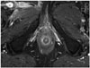
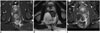
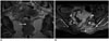
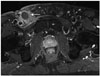
Anal cancers display high signal intensity relative to skeletal muscles on T2-weighted images, but lower than normal ischioanal fat, and low to intermediate signal intensity on T1-weighted images. They are markedly enhanced after intravenous gadolinum contrast injection (Fig. 3) (17,18,19, 29). Anal cancers can extend to the rectum with skip lesions seen at a distance from the primary site, involving low or even middle rectum. They may develop on condylomas, particularly in HIV and HPV positive patients (Figs. 4, 5).
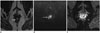
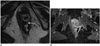
Radiologists should be aware of the possibility of neoplastic changes in long-standing anorectal fistulas, and should be alerted by the presence of soft tissue within the fistula (30). In such cases, AC will generally display T2-weighted high signal intensity, due to colloid content and mesh like internal enhancement (Fig. 6).
Anal melanoma is rare, accounting for only 2% of all melanomas (31), and is generally homogeneous. Primary anal lymphomas are also very rare, and appear as a mass with focal and/or circumferential thickening of the anal canal, potentially extending into the rectum, mildly enhancing after contrast medium injection, and typically without luminal obstruction (32).
Staging of Anal Tumors
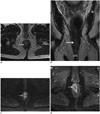
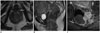
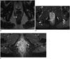
T stage corresponds to the size of the primary tumor (Table 1), assessed by measuring the tumor in its longest diameter on T2-weighted MR images (Figs. 7, 8, 9) (27), but does not take into account the extension to the sphincters. As far as stage T4 is concerned, the ESMO defined that the invasion of the following organs for anal canal carcinoma does not affect T4 staging: external and/or internal sphincter muscles, rectal wall, puborectalis muscle, levator ani muscle and vestibule for tumors originating from the anal margin. For anal margin carcinoma, a T4 lesion is defined by invasion of deeper structures such as the skeletal muscle or cartilage. Unlike N staging for rectal cancer, the location of nodal stations involved determines the N stage for anal cancer (15).
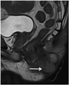
Proximal lymphatic drainage is to perirectal nodes along the inferior mesenteric artery. Infra-dentate and perianal skin tumors drain to the inguinal, femoral and external iliac nodes. Immediately above the dentate line, drainage is to internal pudendal nodes, and to the internal iliac system. All lymph nodes behind the external iliac vessels are considered to belong to the obturator fossa, and are thus a part of the internal iliac group. Nodes along the external and common iliac vessels are considered as distant spread. Criteria for diagnosis of lymph node metastasis are not validated, but enlargement (largest short- axis diameter > 1 cm for mesorectal nodes, and > 1.5 cm for other nodes), heterogeneity or necrosis, irregular contours and strong enhancement are considered to suggest involvement (Fig. 10) (21, 29).
Role of Imaging for Tumor Response Assessment and Detection of Recurrence
Fifty per cent of recurrences occurring within the first 2 years post treatment are located around the primary site of disease, or as pelvic/inguinal lymph nodes (22). ESMO recommends clinical evaluation for complete response at 8 weeks post chemoradiotherapy (CRT), with a 3-to 6-monthly follow up thereafter for a 2-year period, stating that “MRI can capture and document response, but no individual MRI feature appears predictive of eventual outcome” (15). The use of imaging in patients following CRT is still debatable, and there is scarce data in the literature on assessment of tumor response using imaging modalities.
Other international recommendations do not endorse a role of MRI in post CRT surveillance (33, 34). However, these recommendations are based on series of very small sample size (17, 18, 29); in current practice, MRI often serves as the follow-up imaging modality (16, 17, 35). Although consensus criteria for response have not been described so far, reduction in tumor size, as well as in T2-weighted signal intensity of the treated tumor or associated lymphadenopathy, is indicative of response. The optimal timing for therapy assessment has so far not been defined, but size involution seems to be most evident at 6 months post treatment, and complete response has been reported to occur at around 26 weeks (18). Clinicians generally ask for the MRI to be performed 8 weeks after CRT completion, in accordance with what is established for rectal cancers. This time point is likely to be too early, as not all tumors will have achieved complete response, and also since inflammation is superimposed on the treated cancer.
A retrospective study by Kochhar et al. (16), comprising of 74 patients with non-metastatic anal squamous cell carcinoma with a demonstrable anal lesion on baseline staging treated with CRT undergoing 3 and 6 months MRI follow-up, has recently shown that tumor regression grade (TRG) 1/2 scores at 3 and 6 months had a 100% negative predictive value, and that TRG 4/5 scores at 6 months had a 100% positive predictive value. However, no imaging characteristic or TRG score independently prognosticated for late relapse or 3-year disease free survival (16). Reduction in size was seen at 3 (mean 28.79%) and 6 months (mean 81.39%), and reduction in signal intensity was recorded in 69% and 84% of patients at 3 and 6 months, respectively. Nodal disease was downstaged in 33/36 patients, with N0 status in 32 of these patients. The benefit of FDG-PET/CT studies seems to be detection of residual sub-clinical pelvic or extra-pelvic/para-aortic node involvement, rather than assessment of tumor response (13).
Complications Post-CRT
Radiation proctitis as well as fistula (Fig. 11) and abscesses may complicate radiotherapy. Radionecrosis is an uncommon event occurring in up to 10% of patients undergoing radiotherapy for anal cancer (36). It causes clinical (pain, anal stenosis, mucositis and diarrhea) and diagnostic problems. MRI finding of a hypointense area on T1/T2-weighted MR images with only a peripheral enhancement rim, is suggestive of this diagnosis (Fig. 12).
MRI of Recurrent Tumors
Recurrent disease is defined as initial complete response to therapy, with subsequent positive biopsies, more than six months after completion of treatment. Patients with recurrent disease may benefit from surgical salvage (13), and MRI is the best post- operative imaging modality. Similar to rectal cancers (37), T2-weightedhypointense signal at the site of the primary tumor after CRT, is a morphological sign consistent with response, as it represents fibrosis. However, MRI is unable to reliably exclude residual neoplastic foci within the fibrosis. Recently, Kochhar et al. (16) have described a novel ‘tram track’ sign on post-CRT scans, defined as parallel linear low signal at the inner and outer margin of the internal sphincter at the site of the original tumor. This sign had a negative predictive value for early local relapse of 83% at 6 months, and was present in more than half the patients (16). CT scan of thorax and abdomen (or PET/CT) is advised to rule out distant metastases. Tumors that are likely to relapse include tumors larger than 4 cm, with nodal involvement, and basaloid subtypes (23).
 XML Download