

 PDF
PDF ePub
ePub Citation
 Print
Print


INTRODUCTION
Transthoracic needle biopsy (TNB) is a common and minimally invasive procedure for patients with focal lung lesions (123). The goal of TNB is to obtain adequate tissue samples for histopathologic diagnosis; however, practitioners may use different types of devices or techniques at each step of the procedure. The advantages and disadvantages of using a certain technique such as a specific imaging modality (456), a specific needle type (67), or a specific method to prevent complications (8) have been studied extensively, typically based on research from a single institution. However, only a few studies (9101112) were based on a nationwide survey of the actual clinical application of the techniques mentioned above.
In this study, we set out to reexamine current practices for TNB because of a few recent changes in the clinical settings or circumstances where it is performed (as follows). First, detection of genetic mutation has emerged as an important part of lung cancer management (13). Second, more patients seemed to be undergoing surgery for early-stage adenocarcinoma without undergoing preoperative TNB, possibly due to increased ability to recognize specific radiologic features of adenocarcinoma (14). Third, cone beam computed tomography (CBCT) was developed several years ago and had replaced other guiding modalities in a few hospitals (15).
The purpose of this study was to reveal the methods and preferences of radiologists in Korea who currently perform TNB.
MATERIALS AND METHODS
This study was approved by the Institutional Review Board. The requirement for informed consent was waived.
An email survey with 71 questions on TNB was sent to all 240 members of the Korean Society of Thoracic Radiology (KSTR) in March 2016. The survey aimed to identify the TNB-related workflow and practice patterns in each hospital and the technical choices made by each practitioner. Most questions were either multiple-choice or yes-no questions (Supplementary in the online-only Data Supplement), and these became the subject of the current data analysis; however, there were 15 subjective questions that were excluded from the current study.
In the first part of the questionnaire, information on the general characteristics of the respondents (name, affiliation, general experience with thoracic imaging, and experience with TNB) were collected. Then, there were questions on the technical aspects of TNB, followed by questions on important TNB-related complications (air embolism, pneumothorax, tension pneumothorax, and pulmonary hemorrhage). Respondents could skip questions if they desired. For respondents with 5 or more years of experience performing TNB (“> 4-year group”), there were questions (asterisked questions in Supplementary in the online-only Data Supplement) on the differences between the current practice and that of 5 years ago.
To facilitate reliable answers, we assured the respondents that their names and affiliations would be collected for the sole purpose of preventing duplicate answers and that those entries would be deleted before data analysis. Responses were received over a period of one month; in addition, reminders were sent out twice during this period. Descriptive statistics of the data from the survey are summarized in this paper.
RESULTS
Q1, Q2, and so on refer to the question number in Supplementary (in the online-only Data Supplement) related to each result.
Basic Characteristics (Q1-Q3, n = 60)
We sent a survey containing 71 questions to 240 KSTR members. Of 240 members in 106 institutes, 60 members (from 42 academic centers and 5 community-based hospitals) finished the survey and responded (response rate of 25%). Forty-two (70%) of the 60 respondents had over 5 years of experience in performing TNB (“> 4-year group”).
Hospitalization, Consultation, and Record Keeping (Q4-Q9, n = 60)
Fifty-four (90%) respondents answered that they perform TNB only in inpatient settings, and five (8.3%) answered that they do it in outpatient settings either occasionally (n = 4) or often (n = 1). Biopsy requests were received via electronic medical records (EMR) (n = 48, 80%), an internet or an intranet platform outside EMR (n = 4, 6.7%), by phone (n = 2, 3.3%), or through personal visits (n = 6, 10%). All respondents appeared to perform at least 80% of the biopsy requests they received. One respondent said that he/she never declines a TNB request; others performed 90-99% (n = 42; 70% of respondents) or 80-89% (n = 17; 28.4% of respondents) of the requested procedures. Almost all respondents answered that TNB is done exclusively by thoracic radiologists, except one who answered that it is done by an interventional radiologist. Approximately half of the respondents said that they kept TNB records with them in the form of offline spreadsheets (n = 27, 45%), paper reports (n = 7, 11.7%), or online spreadsheets (n = 5, 8.3%); 21 respondents (35%) answered that they do not keep separate TNB records. The majority (n = 53, 88.3%) of respondents answered that they regularly audited TNB results.
Indication
Resectable Lung Lesion (Q10-Q11; n = 60 and 41)
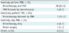
On the question on “TNB in a patient with a focal lung lesion with a high probability of being a resectable-stage lung cancer,” 42 (70%) respondents said that TNB is initially performed, with or without bronchoscopy; 7 (11.7%) said that TNB is selectively done only for inconclusive bronchoscopy results; 5 (8.4%) said that they routinely skip TNB (Table 1). Half of the > 4-year group (46.3% of the 41 who submitted answers) said that this policy has not changed significantly over the past 5 years; 16 (39%) said that the rate of direct surgery (skipping TNB) seems to have increased in the same period.
Subsolid Lesion (Q12, n = 54)
Policies on TNB of subsolid (part-solid or pure ground-glass nodule) lesions varied: 19 (35.2%) respondents never perform TNB, 17 (31.5%) respondents frequently perform TNB, and 17 (31.5%) respondents occasionally perform TNB for subsolid lesions.
High-Risk Patients (Q13, n = 60)
For patients perceived to be at a high risk for procedure-related complications, 27 respondents (45%) said that they occasionally perform TNB; 11 respondents (18.3%) said that they often perform TNB. Only 3 respondents (5%) said that they consistently filter out such cases. Two respondents (3.3%) said that they still perform TNB even if the patient has a high risk for complications. The remaining 17 (28.3%) respondents had no set policy.
Rebiopsy (Q14-Q16; n = 41, 39, and 38, respectively)
The questions were for the > 4-year group only. Out of 41 respondents, 25 (61%) believed that the number of rebiopsy (i.e., TNB in patients with prior histopathologic diagnosis of lung cancer) had increased over the past 5 years. Molecular analysis for an established target therapy (n = 17, 43.6%) and clinical trial of a new drug (n = 11, 28.2%) were listed as the two most common reasons for rebiopsy (39 respondents). A less frequent reason for rebiopsy was suspicion of synchronous cancer based on unresponsiveness to treatment (n = 6, 15.4%). The main reasons for the increase in rebiopsy over the past 5 years were thought to be molecular analysis for an established target therapy (n = 19, 50%) and the clinical trial of a new drug (n = 14, 36.8%).
Biopsy Technique (Q17-Q25: n = 60 for Q17-Q19 and Q22; n = 59 for Q20, Q21, Q25; n = 57 for Q23; n = 56 for Q24)
Coaxial needle was the most preferred tool (n = 33, 55%) followed by non-coaxial cutting needle (n = 19, 31.7%) and aspiration needle (n = 6, 10%). The most popular needle size was 20 G (n = 34, 56.7%) followed by 18 G (n = 21, 35%). The majority (n = 51, 85%) of respondents said that they conducted only one pleural pass; 27 (45%) respondents said that they make multiple needle passes using a coaxial system; 24 (40%) respondents said that they use a non-coaxial system and make only one needle pass. Only 7 (11.7%) respondents said that they routinely make two pleural passes.
Opinions on whether the diameter of a biopsy needle influences the accuracy of histopathologic diagnosis and molecular analysis for lung malignancy varied. Among 59 respondents, 27% thought that it influences neither; 20.3% thought it influences both; 39% said that they used only one diameter of TNB needle and could not answer the question.
The most popular imaging modality was conventional CT (n = 34, 56.7%) followed by fluoroscopy (n = 18, 30%) and CBCT (n = 15, 25%). Among 57 respondents, only 20 respondents had any experience with CT fluoroscopy; 16 respondents reported that they have put a hand within the gantry opening while the X-ray was in operation, and the other 4 respondents said that they have never done so because they use intermittent mode only. On the question about the future introduction of robotic arm in TNB and guiding modality, 36 (64.3%) respondents said that they would choose CT fluoroscopy if robotic arm becomes available; 11 (19.6%) respondents said they would choose CBCT; however, 8 (14.3%) respondents said that they would still use conventional CT.
On the question about TNB with interlobar fissure traversal, 26 (44.1%) respondents said that they pass a needle across the interlobar fissure occasionally when it lies in the most feasible path and regardless of operability; 12 (20.3%) said they use such a path frequently, also regardless of operability; the other 12 (20.3%) said they use such a path exclusively in inoperable patients; 9 (15.3%) said they never do it.
Localization (Q26-Q28; n = 55, 48, and 52, respectively)
Among 55 respondents, 48 (87.3%) had experience in the preoperative localization of lung nodules: hook wire was the most commonly used tool (n = 42, 87.5%) followed by radiopaque markers, such as barium and Lipiodol (n = 18, 37.5%), and dye injection (n = 14, 29.2 %). Only 7 (13.5%) respondents said that they had experience in fiducial marker placement for CyberKnife robotic radiosurgery.
Complications (Q29-Q40)
Out of 60 respondents, 9 (15%) respondents said they had encountered air embolism; 4 (44.4%) respondents used a non-coaxial method at the time of the event; 5 (55.6%) respondents said that the lesion was peripherally located; 6 (66.7%) respondents recalled that the needle did not appear to traverse a visible vessel; the majority recalled that the needle traversed the aerated lung by more than 2 cm (n = 4, 2-5 cm; n = 3, > 5 cm).
Out of 60 respondents, 14 (23.3%) respondents claimed that they had encountered tension pneumothorax with the following evidence: sudden dyspnea (n = 7, 50%), abrupt increase of pneumothorax on CT (n = 4, 28.6%), or drop in blood pressure (n = 2, 14.3%). All 11 respondents said that the most likely risk factor for tension pneumothorax was emphysema.
On the question on post-procedural positioning of a patient with TNB-related pulmonary hemorrhage shown on CT, 27 (45.8%) of 59 respondents said that they always turn patients to a biopsy-side-down decubitus position; 25 (42.4%) said that they turn patients to a biopsy-side-down decubitus position only if there is hemoptysis; others answered that they always place patients in a supine (n = 6, 10.2%) or a sitting (n = 1, 1.7%) position.
DISCUSSION
The survey revealed a high variation in how Korean radiologists perform TNB. There were a few unexpected results as well. For example, we assumed that TNB is performed only in hospitalized patients in Korea, even though TNB is most often performed in an outpatient setting in the United States and British Thoracic Society (BTS) guidelines also state that TNB can be performed as a day-case procedure, except in high risk patients (2). We thought that TNB was entirely an inpatient procedure because the admission fees, which are regulated by the government, are relatively cheap in comparison to per capita income. In addition, some private insurance companies require hospitalization for full reimbursement of procedures like TNB. The results showed that that is not the case and there are occasions where an outpatient procedure is preferred by either the radiologist or the patient.
Transthoracic needle biopsy for a resectable lung lesion is a complicated issue. There is often debate between the referring physicians and the radiologist performing TNB on which policy serves the best interest of the patient. We expect that the information gathered from many different hospitals through this survey will help individual radiologists with this issue. The 2003 American College of Chest Physicians guidelines state “TNB has no role in patients who are candidates for surgical resection” (16); in a Japanese study on epidermal growth factor receptor mutation-positive non-small cell lung cancer patients, TNB accounted for less than 8% of the samples (both for biopsy and cytology) for initial diagnosis (17); therefore, we believe that far too many TNBs are being performed in Korea for resectable lung lesions. For subsolid lesions, policies varied greatly on whether to perform TNB (35.2% who answered “never” vs. 31.5% who answered “frequently”). However, the surgical resection of malignant-looking ground-glass nodule without a preoperative tissue diagnosis has been shown to have no disadvantages compared to the preoperative tissue diagnosis group in terms of recurrence-free survival (14). Unnecessary TNB can result in undesirable results such as procedure-related complications, surgery delays, and increased hospital stays. Fortunately, a significant (39%) number of respondents saw that the trend is changing towards direct surgery for resectable focal lung lesions.
This survey did not investigate the reasons behind the preference for cutting needles and the coaxial technique. However, we believe the popularity of core biopsies is unrelated to the recent increase in personalized medicine because of the following reasons. First, only a few respondents have changed biopsy tools over the past 5 years. This is in contrast to a recent Society of Thoracic Radiology (STR) survey by Lee et al. (12), which showed that the popularity of core needle biopsy has increased considerably over the past decade in the United States. Second, a previous survey by Jin et al. (9) indicated that the coaxial system was already the preferred tool as early as in 2008. Whatever the reason, the fact that core lung biopsy is popular is aligned with the interest of patients, given the increasing number of molecular analysis in lung cancer (1819202122).
According to the respondents, the number of rebiopsy for molecular analysis and clinical trial of a new drug have increased over the past 5 years. Thus, the main role for rebiopsy seems to be shifting from salvaging a failed needle biopsy or a bronchoscopy (2324) toward identifying patients indicated for target therapy or immunotherapy.
In the STR study by Lee et al. (12), most respondents reported making 3 or more needle passes during TNB. We did not directly ask about the exact number of needle passes in our study, but 45% of respondents reported that they make multiple needle passes. Some studies (252627) indicate that at least 3 needle passes appear optimal for histopathologic diagnosis of lung malignancy and mutation study. The current practice of Korean radiologists seems to be consistent with the results of those studies.
In this survey, 90% of respondents used CT-based image guidance: CT (56.7%), CT fluoroscopy (8.3%), or CBCT (25%). Thus, the use of CT guidance has increased since the study by Jin et al. (9) in which 41% of respondents used CT and 10.3% used both CT and fluoroscopy. The STR study by Lee et al. (12) reported an increase in the use of CT guidance compared to 10 years ago (from 70 to 95%); however, the increase was more significant in our study. Notably, a significant number of Korean radiologists use CBCT for guidance while such responses were nonexistent in Lee et al.'s study (12). This result may be due to the difference in popularity of CBCT as an imaging guidance for TNB between countries. The current survey showed that the majority of radiologists avoid using CT fluoroscopy, but they were willing to switch to CT fluoroscopy if robotic arms are made available and the issue of radiation exposure is resolved. The fact that 16 of the 20 respondents who do use CT fluoroscopy in their practices let their own hands be exposed to high-dose X-ray to facilitate real-time monitoring of needle position strongly suggests that the combination of robotic arm and CT fluoroscopy is the way to go.
Radiologists seem quite liberal about traversal of the interlobar fissure during TNB. It seems that they see that the risks associated with vessel traversal (air embolism and hemorrhage) outweigh those associated with traversing an interlobar fissure (pneumothorax and tumor implantation).
Preoperative tumor localization procedures are performed less frequently than TNBs; however, the results show that most respondents have at least some prior experience. There is a large overlap between the techniques used in localization and TNB; therefore, it can be said that patient accessibility to the localization procedure is high in Korea.
The probability of air embolism is very low for a single procedure (0.02% to 0.07%) (282930); however, as many as 15% of our respondents reported that they have encountered it. Contrary to popular belief, the use of the coaxial technique, lesion location, and vessel traversing was not closely related to the occurrence of air embolism in this survey. The lack of correlation between the use of the coaxial technique and air embolism is supported by some air embolism reports that used a non-coaxial method (3031323334). The fact that the length of a traversed aerated lung was greater than 2 cm in 77.8% of the surveyed cases may be important. We suggest that radiologists pay special attention to the occurrence of air embolism when attempting TNB for a deeper target.
There was a substantial difference between the current KSTR survey and the STR survey by Lee et al. (12) with respect to the prevention method for pneumothorax and sedation. We asked about the pneumothorax prevention method (excluded from analysis because it was a subjective question); all 27 respondents said they change the patient position following TNB. However, the prevailing method for pneumothorax prevention among STR members was an autologous blood patch. In addition, the use of hydrogel plugs was recently approved by the FDA for pneumothorax prevention (35). We believe that Korean radiologists should add blood patches and hydrogel plugs in their pallet of pneumothorax-prevention methods. We did not inquire about pre-procedural sedation and local anesthesia, but the study by Lee et al. (12) showed that IV sedation is frequently (43%) used by STR members. This finding merits further investigation because the use of IV sedation has been discouraged by BTS guidelines (2) and is currently uncommon in Korea.
Our study has limitations in that only 25% of KSTR members responded to the survey, and that the number of respondents was relatively small. However, the percentage of the respondents among actual performers may be higher because not all KSTR members perform TNB in their practices. Another limitation is the generalizability of results that are constrained to a survey conducted in a single country.
In conclusion, despite high variation in how TNB is performed in Korea, some patterns were noted. It is common for patients with resectable-stage lung cancer to undergo TNB prior to surgery. Only a small percentage of high-risk cases are actively being filtered out by radiologists. Rebiopsy is now more common than before mainly because of personalized medicine. The most popular type of needle is the coaxial system; the most popular modality for guidance is still CT. We expect that the results of this survey will help enhance communications between the practitioners regarding the procedure.
 XML Download